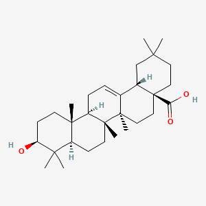
DR ANTHONY MELVIN CRASTO,WorldDrugTracker, helping millions, A 90 % paralysed man in action for you, I am suffering from transverse mylitis and bound to a wheel chair, With death on the horizon, nothing will not stop me except God................DR ANTHONY MELVIN CRASTO Ph.D ( ICT, Mumbai) , INDIA 25Yrs Exp. in the feld of Organic Chemistry,Working for GLENMARK GENERICS at Navi Mumbai, INDIA. Serving chemists around the world. Helping them with websites on Chemistry.Million hits on google, world acclamation from industry, academia, drug authorities for websites, blogs and educational contribution

Thursday 7 August 2014

1-Ethylpropylamine NMR


1-Ethylpropylamine

Formula: C5H13N


IR spectrum

The two bands at 3388 and 3292 indicate a primary amine (-NH2). The bands at 3000-2850 indicate C-H alkane stretches.


C5H13N
Rule 3, omit the N and one H, gives C5H12
5 - 12/2 + 1 = 0 degrees of unsaturation.
No pi bonds or rings.


13 C NMR


................

1H NMR


NMR answer
Note that C is showing as 2 2H peaks, instead of a 4H peak. This may be caused by a similar effect as in Problem 4 - on each carbon C, the H closer to the N will be in a slightly different environment. If the rotation of this carbon is hindered, these Hs may have a different chemical shift.


...............

Take a tour   Indian food
North Indian Food vahrehvah.com
SAMOSA
.
THALI
/////////

Wednesday 6 August 2014

3 Isobutylglutaric acid Spectral data


3-isobutylglutaric acid
3-(2-Methylpropyl)pentanedioic Acid;
Pentanedioic acid,3-(2-Methylpropyl)-
Purified 3-isobutylglutaric acid is a solid with a melting point in the range of about 40°C to about 42°C.

1H NMR (CDC13 200 MHz) :
δ 0.92 (d, 6H, J = 6.6 Hz) ,
1.23 (dd, 2H, Jχ = 6.6 Hz, J2 = 6.5 Hz) ,
1.64 ( , 1 H) ,
2.25-2.40 (m, 1 H) ,
2.40-2.55 (m, 4 H) .

13C NMR (CDCI3) :
δ 22.4,
25.1,
29.5,
38.4,
43.4,
179.2, two carbonyls

IR (KBr) : 680.7, 906.4, 919.9, 1116.6, 1211.1, 1232.3, 1249.6, 1301.7,
1409.7, 1417.4, 1448.3, 1463.7, 1704.8, 2958.3, 3047.0 cm"1.

......................................................................................................
ARKIVOC 2010 (x) 266-275 
IR (cm-1): 3436, 2953, 1575;
1H NMR (D2O, δ ppm):
0.70 (d, 6H, CH3, J 6.0 Hz),
0.97 (s, 2H, CH2), 1.49 (s, 1H, CH), 1.95 (s, 4H, CH2), 2.05 (s, 1H, CH);
13C NMR (D2O, ppm):
182.89, 43.66, 31.90, 24.60, 22.27;
MS: m/z: 187 (M-H+);
Analysis calcd. for C9H16NO4:
C,57.43; H, 8.57% Found: C, 57.41; H, 8.55%.

......................................
Example 1
To a four neck round bottom flask fitted with a mechanical stirrer, condenser and charging tube, was charged isovaleraldehyde (1.0 kg, 11.61 mole), cyclohexane (1.35 L), ethyl cynoacetate (1.28 kg, 11.38 mole) and di-n-propylamine (11.74 g). The reaction mass was heated to reflux and water was removed azeotropically. After complete removal of water (˜208 ml), cyclohexane was distilled from the reaction mass followed by removal of traces of cyclohexane under vacuum. The reaction mass was cooled to 30-35° C. and diethyl malonate (2.027 kg, 12.67 mole) was added followed by addition of di-n-propylamine (106.91 g). The reaction mass was heated to 50-55° C. for 3-5 hours and then cooled to 25-30° C. Then hydrobromic acid (47%, 23.76 L) was added and the mass was refluxed at 100-125° C. for 6-10 hours. The reaction mass was cooled to 25-30° C. and extracted with toluene. The toluene was distilled off to obtain 3-isobutylglutaric acid in a yield of 1.54 kg (71%) having GC purity of 93.59%.
 NMR ASSIST
USE SPECTRUM OF GLUTARIC ACID TO UNDERSTAND ABOVE COMPD
GLUTARIC ACID
IR


1H NMR 
spectrum for Glutaric acid


13 C NMR
spectrum for Glutaric acid


2D [1H,1H]-TOCSY
spectrum for Glutaric acid

Tuesday 5 August 2014

Travoprost spectral data


Travoprost structure.svg


Travoprost
cas 157283-68-6
[1R-[lα(Z),2β(lE,3R*),3α,5α]]-7-[3,5-Dihydroxy-2-[3-hydroxy-4-[3-(trifluoromethyl)phenoxy]-1 -butenyl]cyclopentyl]-5-heptenoic acid, 1 -methylethylester
(+)-16-m-trifluoromethylphenoxy tetranor Prostaglandin F isopropyl ester; (+)-Fluprostenol ispopropyl ester
(+)-(5Z,9α,1α,13E,15R)-trihydroxy-16-(3-(trifluoromethyl)phenoxy)-17,18,19,20-tetranor-prosta-5,13-dien-1-oic acid, isopropyl ester
(+) – Fluprostenol isopropyl ester,
CAS Name: (5Z)-7-[(1R,2R,3R,5S)-3,5-Dihydroxy-2-[(1E,3R)-3-hydroxy-4-[3-(trifluoromethyl)phenoxy]-1-butenyl]cyclopentyl]-5-heptenoic acid 1-methylethyl ester
Additional Names: (+)-16-[3-(trifluoromethyl)phenoxy]-17,18,19,20-tetranorprostaglandin F2a isopropyl ester; (+)-9a,11a,15-trihydroxy-16-(3-trifluoromethylphenoxy)-17,18,19,20-tetranor-5-cis-13-trans-prostadienoic acid isopropyl ester
Manufacturers’ Codes: AL-6221
Trademarks: Travatan (Alcon)
Percent Composition: C 62.39%, H 7.05%, F 11.39%, O 19.18%
Travatan, Travatan Z, AL-6221, Travatanz, Travatan Alcon, Travatan (TN), Travatan, Travoprost, Travoprost [USAN]
Molecular Formula: C26H35F3O6
Molecular Weight: 500.54771
Alcon (Originator)
Antiglaucoma Agents, OCULAR MEDICATIONS, Ophthalmic Drugs, Prostaglandins, Prostanoid FP Agonists
Properties: Colorless oil. [a]D20 +14.6° (c = 1.0 in methylene chloride). Very sol in acetonitrile, methanol, octanol, chloroform. Practically insol in water.
Optical Rotation: [a]D20 +14.6° (c = 1.0 in methylene chloride)
Therap-Cat: Antiglaucoma.
Ophthalmic solution used for the reduction of elevated intraocular pressure in patients with open-angle glaucoma or ocular hypertension who are intolerant of other intraocular pressure lowering medications or insufficiently responsive (failed to achieve target IOP determined after multiple measurements over time) to another intraocular pressure lowering medication.

read at


Org Process Res Dev2002,6, (2): 138
 (5Z,13E)-(9S,11R,15R)-9,11,15-Trihydroxy-16-(m-trifluoromethylphenoxy-17,18,19,20-tetranor-5,13-prostadienoic Acid, Isopropyl Ester (2).
The silyl-protected compound (20a+b) (202 g, 277 mmol) ………..DELETED……………………………………… All relevant fractions were combined and concentrated to give the title compound 2 (97 g, 70%) as a colourless oil,

  +14.6 (c 1.0, CH2Cl2);

 IR νmax (film) 3374 and 1727 cm-1

;1H NMR (400 MHz, CDCl3
δ 7.39 (1H, t, J = 8), 7.22 (1H, d, J = 8), 7.15 (1H, s), 7.08 (1H, d, J = 8), 5.70 (2H, m), 5.40 (2H, m), 4.98 (1H, heptet, J = 6.5), 4.52 (1H, m), 4.18 (1H, m), 3.97 (3H, m), 3.25 (2H, br s), 2.60 (1H, br s), 2.38 (1H, m), 2.30−1.96 (7H, m), 1.76 (1H, dd, J = 16, 4), 1.65 (2H, quintet, J = 7), 1.55 (1H, m), and 1.20 (6H, d, J = 6); 


13C NMR (100 MHz, CDCl3
δ 173.57, 158.67, 135.45, 131.87 (q, J = 32), 130.02, 129.85, 129.75, 128.93, 123.89 (q, J = 270), 118.06, 117.82, 111.48, 77.77, 72.70, 71.99, 70.86, 67.72, 55.82, 50.24, 42.84, 34.00, 26.60, 25.48, 24.83, and 21.81; m/z (CI) 501 (MH+, 21), 321 (34), 303 (44), and 249 (100).


Saturday 2 August 2014

BIMATOPROST SPECTRAL DATA

Bimatoprost.svg
BIMATOPROST
155206-00-1
Lumigan, Latisse, AGN 192024, bimatoprostum, UNII-QXS94885MZ, Lumigan (TN), CHEBI:51230, AC1NSJUW, AGN-192024
Molecular Formula: C25H37NO4
 Molecular Weight: 415.56558
(Zanoni, G. et al., Tetrahedron 66, 7472-7478 (2010); Gutman, A. et al., US 20090163596 (2009)).

SYN
http://newdrugapprovals.org/2014/07/31/bimatoprost/


http://www.google.com/patents/EP2454227A1?cl=en

 BIMATOPROST97 (82.6 mg, 41% over 2 steps) as a clear, colourless oil. Analytical data consistent with the literature (Zanoni, G. et al., Tetrahedron 66, 7472-7478 (2010); Gutman, A. et al., US 20090163596 (2009)).

max (filmVcm-1 3300 (broad), 2930, 1643, 1550, 1453, 1332, 1293, 1048, 1029, 968, 729, 698
*H NMR (400 MHz; CDCI3) δΗ = 1.09 (t, J = 7.1 Hz, 3H, CH3), 1.42-2.40 (m, 14H, 6 x CH2, 2 x CH), 2.67 (m, 2H, CH2), 3.22 (dq, J = 7.1, 6.3 Hz, 2H, CH2NH), 3.41 (broad s, 3H, 3 x OH), 3.80-4.30 (broad m, 3H, 3 x CHOH), 5.37 (m, 2H, 2 x =CH), 5.47 (dd, J = 15.2, 7.9 Hz, 1H, =CH), 5.59 (dd, J = 15.2, 7.9 Hz, 1H, =CH), 5.90 (broad s, 1H, NH), 7.17 (m, 3H, ArCH’s), 7.26 (m, 2H, ArCH’s)
13C NMR ( 100 MHz; CDCI3) 5C = 14.8 (CH3), 25.4 (CH2), 25.6 (CH2), 26.7 (CH2), 31.9 (CH2), 34.4 (CH2NH), 35.8 (CH2C=0), 38.8 (CH2), 42.9 (CH2), 50.2 (CH), 55.5 (CH), 72.3 (CHOH), 72.4 (CHOH), 77.7 (CHOH), 125.8 (ArCH), 128.4 (2 x ArCH), 128.5 (2 x ArCH), 129.1 (=CH), 129.7 (=CH), 133.7 (=CH), 135.1 (=CH), 142.0 (ArC), 173.4 (C=0)
m/z (ESI+) 438.2 [MNa]+
HRMS (ESI+) calcd for Q^IV^Na [MNa]+ 438.2614, found 438.2615
[a]D 22 +41.1 (c. 0.35, CH2CI2) (lit. – Zanoni, G. et al., Tetrahedron 66, 7472-7478 (2010), +32.7 (c. 0.33, CH2CI2)) (lit. – Gutman, A. et al., US 20090163596 (2009), +36 (c. 1, MeOH))

LATANOPROST SPECTRAL DATA

Latanoprost.svg
Latanoprost
isopropyl-(Z)7[(1R,2R,3R,5S)3,5-dihydroxy-2-[(3R)-3-hydroxy-5-phenylpentyl]cyclopentyl]-5-heptenoate.
130209-82-4
XA41, PhXA34 [as 15 (R, S) -isomer], PhXA41, Xalatan
(Zanoni, G. et al., Tetrahedron 2010, 66, 7472)
SYNTHESIS
http://newdrugapprovals.org/2014/08/02/latanoprost/


http://www.google.com/patents/WO2013186550A1?cl=en

latanoprost 77 (71 mg, 64 %) as a clear colourless oil. The IR, 13C, and optical rotation data were consistent with the literature (Zanoni, G. et al., Tetrahedron 2010, 66, 7472). Rf = 0.44 (EtOAc)
vmax (neatVcm“1 3360 (broad), 2980, 2931, 2857, 1712, 1495, 1454, 1374, 1311, 1247, 1180, 1106, 1030, 966, 910, 820, 731, 699
*H NMR (400 MHz; CDCI3) δΗ = 1.23 (6 H, d, J = 6.4 Hz, 2 x CH3), 1.30-1.90, (14 H, m, 5 x CH2, 2 x CH, 2 x OH), 2.07-2.39 (6 H, m, 3 x CH2), 2.45 (1 H, d, J = 5.5 Hz, OH), 2.63- 2.86 (2 H, m, CH2), 3.68 (1 H, br.s, CHO ), 3.95 (1 H, br.s, CHOH), 4.18 (1 H, br.s, CHO ), 5.01 (1 H, sept., J = 6.4 Hz, OCH(CH3)2), 5.35-5.52 (2 H, m, 2 x =CH), 7.16-7.24 (3 H, m, ArH’s), 7.25-7.32 (2 H, m, ArH’s)
13C NMR (125 MHz; CDCI3) 5C = 21.9 (2 x CH3), 24.9 (CH2), 26.6 (CH2), 26.8 (CH2), 29.6 (CH2), 32.1 (CH2), 34.0 (CH2), 35.7 (CH2), 39.0 (CH2), 42.5 (CH2), 51.8 (CH), 52.7 (CH), 67.6 (OCH), 71.2 (OCH), 74.5 (OCH), 78.6 (OCH), 125.7 (CH), 128.3 (2 x ArCH), 128.3 (2 x ArCH), 129.3 (CH), 129.5 (CH), 141.1 (ArC), 173.5 (C=0)
[a]D 23 33.0 (c. 1.0, MeCN) (lit, [a]D 20 32.7 (c. 1.0, MeCN))


...............

Take a tour   Indian food
North Indian Food vahrehvah.com
SAMOSA
.
THALI
/////////


Friday 1 August 2014

Decursinol angelate spectral data and interpretation


Decursinol angelate
C19H20O5
328.36

MASS


EIMS m/z 328 [M]+


1H NMR

(300MHz, CDCl3) δ 1.31, 1.33 (3H, s, gem(CH3)2), 1.77 (3H, s, 4"-H), 1.80 (3H, d, J=7.8, 5"-H), 2.83 (dd, J=17.4, 4.8, 4'-H), 3.18(1H, dd, J=17.4, 4.8, 4'-H), 5.05 (t, J=5.0, 1H, 3'-H), 6.02 (1H, q, 6.16 (1H, d, J=9.6, 3-H), 6.72 (1H, s, 5-H), 7.08 (1H, s, 8-H), 7.53 (1H, d, J=9.6, 4-H)

broad line below is  ''


difficulty, then use the numbering in below fig


13 C NMR

(100MHz, CDCl3) δ 15.7 (C-4"), 20.5 (C-5"), 23.2, 25.0 (gem(CH3)2), 27.8 (C-4'), 70.0 (C-3'), 76.6 (C-2'), 104.6 (C-8), 112.8 (C-10), 113.2 (C-3), 115.8 (C-6), 127.3 (C-2"), 128.6 (C-5), 139.4 (C-3"), 143.1 (C-4), 154.2 (C-9), 156.4 (C-7), 161.2 (C-2) 167.0 (C-1")


HPLC
http://www.phcog.com/article.asp?issn=0973-1296;year=2014;volume=10;issue=37;spage=34;epage=39;aulast=Hwang


Oleanolic acid spectral data and interpretation


Chemical structure for Oleanolic Acid


Oleanolic acid

Oleanolic acid
(4aS,6aR,6aS,6bR,8aR,10S,12aR,14bS)-10-hydroxy-2,2,6a,6b,9,9,12a-heptamethyl-1,3,4,5,6,6a,7,8,8a,10,11,12,13,14b-tetradecahydropicene-4a-carboxylic acid

Oleanic acid, Caryophyllin, Astrantiagenin C, Giganteumgenin C, Virgaureagenin B, 3beta-Hydroxyolean-12-en-28-oic acid, OLEANOLIC_ACID
Molecular Formula: C30H48O3   
Molecular Weight: 456.70032  


Ursolic acid [(3b)-3-Hydroxyurs-12-en-28-oic acid] rarely occurs without its isomer oleanolic acid [(3b)-3-Hydroxyolean-12-en-28-oic acid] They may occur in their free acid form, as shown in Figure 1, or as aglycones for triterpenoid saponins which are comprised of a triterpenoid aglycone linked to one or more sugar moieties. Ursolic and oleanolic acids are similar in pharmacological activity

A pentacyclic triterpene that occurs widely in many PLANTS as the free acid or the aglycone for many SAPONINS. It is biosynthesized from lupane. It can rearrange to the isomer, ursolic acid, or be oxidized to taraxasterol and amyrin.   


MS
EIMS m/z (rel. int.) 456 [M]+ (5), 412 (3), 248 (100), 203 (50), 167 (25), 44 (51)


IR KBR
(KBr) 3500, 2950, 2850, 1715; 1H-NMR (250 MHz, pyridine-d5) δ: 5.49 (1H, s, H-12), 3.47 (1H, t, J = 8.0 Hz, H-3), 3.30 (1H, m, H-18), 1.12 (3H, s, CH3-27), 0.96 (3H, s, CH3-30), 0.91 (3H, s, CH3-25), 0.89 (3H, s, CH3-23), 0.87 (3H, s, CH3-24), 0.75 (3H, s, CH3-26)


1H NMR
(250 MHz, pyridine-d5)

δ: 5.49 (1H, s, H-12), 3.47 (1H, t, J = 8.0 Hz, H-3), 3.30 (1H, m, H-18), 1.12 (3H, s, CH3-27), 0.96 (3H, s, CH3-30), 0.91 (3H, s, CH3-25), 0.89 (3H, s, CH3-23), 0.87 (3H, s, CH3-24), 0.75 (3H, s, CH3-26)


13 C NMR
(63 MHz, pyridine-d5) δ: 180.2 (C-28), 144.8 (C-13), 122.5 (C-12), 78.0 (C-3), 55.7 (C-5), 48.0 (C-9), 46.6 (C-8, 17), 42.1 (C-14), 39.7 (C-4), 39.4 (C-1), 37.3 (C-10), 33.2 (C-7), 32.9 (C-29), 32.4 (C-21), 30.9 (C-20), 28.7 (C-23), 27.2 (C-2), 26.9 (C-15), 26.1 (C-30), 23.7 (C-11), 23.6 (C-16), 18.7 (C-6), 17.4 (C-26), 16.5 (C-24), 15.5 (C-25)


http://www.google.com/patents/US20120237629

FIG. 4 shows the 1H NMR spectrum of oleanolic acid;
FIG. 5 shows the 13C NMR spectrum of oleanolic acid;
FIG. 6 shows the 13C DEPT NMR spectrum of oleanolic acid;
FIG. 7 shows the 113C HSQC NMR spectrum of oleanolic acid;

see below


EXAMPLE 2 Extraction and Isolation of Oleanolic Acid (9) and Maslinic Acid (10) from Cloves
Syzygium aromaticum dried buds or whole cloves were obtained commercially. The cloves (1.5 kg, whole) of Syzygium aromaticum were sequentially and exhaustively extracted with hexane and ethyl acetate to give, after solvent removal in vacuo, a hexane extract (68.8 g, 4.9%) and an ethyl acetate extract (34.1 g, 2.3%). A portion of the ethyl acetate extract (10.0 g), was subjected to chromatographic separation on silica gel (60-120 mesh) column (40×5.0 cm). Elution with hexane/ethyl acetate solvent mixtures (8:2→6:4) afforded pure oleanolic acid (9) (4.7 g, 1.06%), a mixture of oleanolic acid (9) and maslinic acid (10) (0.5 g), and pure maslinic acid (10) (0.25 g). The structures of oleanolic acid (9) and maslinic acid (10) (as 2,3-diacetoxyoleanolic acid) were confirmed by spectroscopic data analysis (1D and 2D 1H NMR and 13C NMR experiments) (FIGS. 4-7 and FIGS. 8-10, respectively).