DR ANTHONY MELVIN CRASTO,WorldDrugTracker, helping millions, A 90 % paralysed man in action for you, I am suffering from transverse mylitis and bound to a wheel chair, With death on the horizon, nothing will not stop me except God................DR ANTHONY MELVIN CRASTO Ph.D ( ICT, Mumbai) , INDIA 25Yrs Exp. in the feld of Organic Chemistry,Working for GLENMARK GENERICS at Navi Mumbai, INDIA. Serving chemists around the world. Helping them with websites on Chemistry.Million hits on google, world acclamation from industry, academia, drug authorities for websites, blogs and educational contribution

Sunday, 12 July 2015

Trimyristin

Trimyristin

Trimyristin

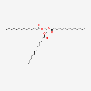
Trimyristin is an ester with the chemical formula C45H86O6. It is a saturated fat which is the triglyceride of myristic acid. Trimyristin is a white to yellowish-gray solid that is insoluble in water, but soluble in ethanol, benzene, chloroform, dichloromethane, and ether.

Name Trimyristin
Synonyms Glycerol trimyristate
Name in Chemical Abstracts Tetradecanoic acid, 1,2,3-propanetriyl ester
CAS No 555-45-3
EINECS No 209-099-7
Molecular formula C45H86O6
Molecular mass 723.18
SMILES code CCCCCCCCCCCCCC(=O)OCC(OC(=O)CCCCCCCCCCCCC)COC(=O)CCCCCCCCCCCCC
Nutmeg

reacts to

Trimyristin
TRIMYRISTIN.png

Occurrence

Trimyristin is found naturally in many vegetable fats and oils.

Isolation from nutmeg

Seed of nutmeg contains trimyristin
The isolation of trimyristin from powdered nutmeg is a common introductory-level college organic chemistry experiment. It is an uncommonly simple natural product extraction because nutmeg oil generally consists of over eighty percent trimyristin. Trimyristin makes up between 20-25% of the overall mass of dried, ground nutmeg. Separation is generally carried out by steam distillation and purification uses extraction from ether followed by distillation or rotary evaporation to remove the volatile solvent. The extraction of trimyristin can also be done with diethyl ether at room temperature, due to its high solubility in the ether. The experiment is frequently included in curricula, both for its relative ease and to provide instruction in these techniques.

1H-NMR

1H NMR
1H-NMR: Trimyristin
300 MHz, CDCl3
delta [ppm] mult. atoms assignment
0.90 m 9 H 14-H (CH3)
1.2-1.4 m 60 H 4-13-H (CH2)
1.5-1.7 m 6 H 3-H
2.33 m 6 H 2-H
4.16 dd 2 H glycerol-C1-Ha
4.31 dd 2 H glycerol-C1-Hb
5.28 m 1 H glycerol-C2-H
7.26

CHCl3
2.11

acetone (impurity)

Isolation of trimyristin from nutmeg

Reaction type: isolation of natural products
Substance classes: carboxylic acid ester, triglyceride, natural product
Techniques: extracting with Soxhlet extractor, evaporating with rotary evaporator, recrystallizing, filtering, heating under reflux, heating with oil bath, stirring with magnetic stir bar
Degree of difficulty: Easy
Batch scale: 25 g Nutmeg

Reaction……….http://kriemhild.uft.uni-bremen.de/nop/en/instructions/pdf/1021_en.pdf

The reaction apparatus consists of a 250 mL round-bottom flask with a magnetic stir bar and a 100 mL soxhlet extraction unit with a reflux condenser. 25 g of finely ground nutmeg are placed into the extraction sleeve and covered with a little glass wool. 150 mL tert-butyl methyl ether are placed into the flask and whilst stirring, the solvent is heated to reflux until the solvent leaving the extraction sleeve is colourless (approximately 5 hours).

Work up

The solvent is evaporated with a final pressure of 20 hPa. The flask containing the residue is cooled in an ice bath or the refrigerator until the contents has crystallized to a thick slurry.
Crude product yield: 12 g;
The crude product is recrystallized from the minimum amount of ethanol. Prior to filtering the crystals, the flask is placed into the refrigerator for at least 30 minutes. The crystalline slurry is filtered and the product is dried in an evacuated desiccator over silica gel. Should the crystals not be colourless after the first recrystallization, a second recrystallization is carried out.
Yield: 6.5 g; melting point 54-55 °C;

Duration of the experiment

Without recrystallization 6 hours

Where can I stop the experiment?

Before and after the evaporation of the solvent

Recycling

The evaporated tert-butyl methyl ether and the evaporated ethanol from the mother liquor are collected and redistilled.

Suggestions for waste disposal

Waste Disposal
residue from mother liquor domestic waste
residue from extraction domestic waste

Operating scheme

Operating scheme

Substances required

Batch scale: 25 g Nutmeg
Educts
Amount Risk Safety
Nutmeg

25 g R S
Solvents
Amount Risk Safety
Ethanol
F F
~ 150 mL R 11 S 2-7-16
tert-Butyl methyl ether
F F Xi Xi
150 mL R 11-38 S 2-9-16-24
Others
Amount Risk Safety
Iodine
Xn Xn N N
0.1 g R 20/21-50 S 2-23.2-23.4-25-61
Solvents for analysis
Amount Risk Safety
Cyclohexane
F F Xn Xn N N
? R 11-38-50/53-65-67 S 2-9-16-33-60-61-62
Acetic acid ethyl ester
F F Xi Xi
? R 11-36-66-67 S 2-16-26-33

Substances produced

Batch scale: 25 g Nutmeg
Products
Amount Risk Safety
Trimyristin

6.5 g R S

Equipment

Batch scale: 25 g Nutmeg
round bottom flask 250 mL round bottom flask 250 mL
Soxhlet extractor 100 mL Soxhlet extractor 100 mL
glass wool glass wool
extraction cone extraction cone
heatable magnetic stirrer with magnetic stir bar heatable magnetic stirrer with magnetic stir bar
oil bath oil bath
reflux condenser reflux condenser
rotary evaporator rotary evaporator
ice bath ice bath
exsiccator with drying agent exsiccator with drying agent
suction filter suction filter
suction flask suction flask

Simple evaluation indices

Batch scale: 25 g Nutmeg
Atom economy
not defined
Yield
not defined
Target product mass
6.5 g
Sum of input masses
250 g
Mass efficiency
26 mg/g
Mass index
39 g input / g product
E factor
38 g waste / g product
Energy input
1500 kJ
Energy efficiency
4.3 mg/kJ

Chromatogram

crude product chromatogram
TLC: crude product
TLC layer Polygram SilG/UV precoated TLC layer; 0.2 mm; silica gel; Macherey & Nagel
mobile phase cyclohexane / EtOAc = 95 : 5
staining reagent Vaughn’s reagent or iodine vapor
Rf (product) 0.51

13C-NMR

13C NMR
13C-NMR: Trimyristin
300 MHz, CDCl3
delta [ppm] assignment
14.08 C14
22.66 C13
24.85-24.89 C3, C17
29.06-31.90 C4-C12
34.04-34.2 C2
62.08 glycerol-C1
68.85 glycerol-C2
172.85 C15
173.26 C1
76.5-77.5 CDCl3


IR

IR
IR: Trimyristin
[KBr, T%, cm-1]
[cm-1] assignment
2950-2850 aliph. C-H valence
1730 C=O valence, ester


Trimyristin[1]
Skeletal formula of trimyristin
Ball-and-stick model of trimyristin
Space-filling model of trimyristin
Names
IUPAC name
1,3-Di(tetradecanoyloxy)propan-2-yl tetradecanoate
Other names
Glyceryl trimyristate; Glycerol tritetradecanoate;[2] 1,2,3-Tritetradecanoylglycerol[3]
Identifiers
555-45-3 Yes
ChemSpider 10675 
EC number 209-099-7
Jmol-3D images Image
PubChem 11148
UNII 18L31PSR28 Yes
Properties
C45H86O6
Molar mass 723.18 g·mol−1
Appearance White-yellowish gray solid
Odor Nutmeg-like
Density 0.862 g/cm3 (20 °C)[4]
0.8848 g/cm3 (60 °C)[2]
Melting point 56–57 °C (133–135 °F; 329–330 K)
Boiling point 311 °C (592 °F; 584 K)
Solubility Slighty soluble in alcohol, ligroin
Soluble in (C2H5)2O, acetone, C6H6,[2] CH2Cl2, CHCl3
1.4428 (60 °C)[2]
Structure
Triclinic (β-form)[3]
P1 (β-form)[3]
a = 12.0626 Å, b = 41.714 Å, c = 5.4588 Å (β-form)[3]
α = 73.888°, β = 100.408°, γ = 118.274°
Thermochemistry
1013.6 J/mol·K (β-form, 261.9 K)
1555.2 J/mol·K (331.5 K)[5][6]
1246 J/mol·K (liquid)[6]
−2355 kJ/mol[6]
27643.7 kJ/mol[6]
Hazards
NFPA 704
Flammability code 0: Will not burn. E.g., water Health code 1: Exposure would cause irritation but only minor residual injury. E.g., turpentine Reactivity code 0: Normally stable, even under fire exposure conditions, and is not reactive with water. E.g., liquid nitrogen Special hazards (white): no code NFPA 704 four-colored diamond
0
1
0
Flash point > 110 °C (230 °F; 383 K)[7]
421.1 °C (790.0 °F; 694.2 K)[7]
Except where otherwise noted, data are given for materials in their standard state (at 25 °C [77 °F], 100 kPa).
    1. References

    2. Merck Index, 11th Edition, 9638.

    3. Lide, David R., ed. (2009). CRC Handbook of Chemistry and Physics (90th ed.). Boca Raton, Florida: CRC Press. ISBN 978-1-4200-9084-0.

    4. Van Langevelde, A.; Peschar, R.; Schenk, H. (2001). “Structure of β-trimyristin and β-tristearin from high-resolution X-ray powder diffraction data”. Acta Crystallographica Section B Structural Science 57 (3): 372. doi:10.1107/S0108768100019121. edit

    5. Sharma, Someshower Dutt; Kitano, Hiroaki; Sagara, Kazunobu (2004). “Phase Change Materials for Low Temperature Solar Thermal Applications” (PDF). http://www.eng.mie-u.ac.jp. Mie University. Retrieved 2014-06-19.

    6. Charbonnet, G. H.; Singleton, W. S. (1947). “Thermal properties of fats and oils”. Journal of the American Oil Chemists Society 24 (5): 140. doi:10.1007/BF02643296. edit

    7. Trimyristin in Linstrom, P.J.; Mallard, W.G. (eds.) NIST Chemistry WebBook, NIST Standard Reference Database Number 69. National Institute of Standards and Technology, Gaithersburg MD. http://webbook.nist.gov (retrieved 2014-06-19)

“MSDS of Trimyristin”
      .
http://www.fishersci.ca
      . Fisher Scientific. Retrieved 2014-06-19.


DRUG APPROVALS BY DR ANTHONY MELVIN CRASTO …..FOR BLOG HOME CLICK HERE
Join me on Linkedin

View Anthony Melvin Crasto Ph.D's profile on LinkedIn

Join me on Facebook FACEBOOK
Join me on twitterFollow amcrasto on Twitter
Join me on google plus Googleplus

 amcrasto@gmail.com

09b37-misc2b027LIONEL MY SON
He was only in first standard in school when I was hit by a deadly one in a million spine stroke called acute transverse mylitis, it made me 90% paralysed and bound to a wheel chair, Now I keep him as my source of inspiration and helping millions, thanks to millions of my readers who keep me going and help me to keep my son happy


…………
Google+

ANTHONY MELVIN CRASTO

Mass Spectrometry: Isotope Effects


Mass Spectrometry: Isotope Effects


This blogpost focuses on the effects of isotopes on mass spectra. It also addresses practical applications of isotope measurement using a mass spectrometer.

Introduction

The ability of a mass spectrometer to distinguish different isotopes is one of the reasons why mass spectrometry is such a powerful technique.The presence of isotopes – a presence that is ubiquitous in nature – gives each fragment a characteristic series of peaks with different intensities.These intensities can be predicted based on the abundance of each isotope in nature, and the relative peak heights can also be used to assist in the deduction of the empirical formula of the molecule being analyzed.

Isotopes 

An ‘isotope’ of any given element is an atom with the same number of protons but a different number of neutrons, resulting in a different overall mass.  Almost all elements have a variety of naturally occurring isotopes – some notable exceptions are fluorine, phosphorous, sodium, and iodine.  For example, Carbon has 6 protons.  In its most common isotope, it also has 6 neutrons, with a total number of protons +neutrons of 12 (called the ‘Mass Number’).  This carbon atom, with a mass number of 12, is written: 12C.  Carbon has two other naturally occurring isotopes, 13C and 14C, which have 7 and 8 neutrons, respectively.  Table 1 below gives the most abundant naturally occurring isotopes for a variety of important elements.  The abundance for secondary isotopes is reported in number of atoms of secondary isotope for every 100 atoms of the most abundant isotope1.

Element
Most Abundant Isotope
Secondary Isotope
Abundance/100 atoms of Primary Isotope
Hydrogen
1H
2H
0.015
Carbon
12C
13C
1.080
Nitrogen
14N
15N
0.370
Oxygen
16O
17O
0.040
18O
0.200
Sulfur
32S
33S
0.800
34S
4.400
Chlorine
35Cl
37Cl
32.50
Bromine
79Br
81Br
98.00
Silicon
28Si
29Si
5.100
30Si
3.400
Table 1. Abundance of several naturally occurring biologically important isotopes1.


Qualitative Peak Analysis

Mass spectrometry distinguishes elements based on a mass to charge ratio, m/z.  Because of this, isotopes play an important role in mass spectra.  Each isotope will show up as a separate line in any mass spectrum with good enough resolution.The Y-axis on a mass spectrum is relative intensity.Therefore, the height of each of the peaks will correspond to the relative abundance of each isotope in the sample.  As an example, examine Figure 1.This is a mass spectrum of a natural sample of atomic carbon.  12C, with a natural abundance of 98.89%, naturally has a very high peak.  13C, with a natural abundance of only 1.11%, has a very low peak.  The radioactive 14C has such a low natural abundance that it is not even seen in relation to the other two carbon isotopes2.  The 12C peak in this spectrum would be called the Base Peak, and would be labeled M+.  The base peak is the largest peak in a spectrum, and the intensity of every other peak is reported in comparison to the base peak.  The 13C peak, with an atomic mass of 1 greater than 12C, would be labeled (M+1)+14C, if it were present, would be (M+2)+
MSIE-Fig1.jpg
Figure 1. Mass spectrum for atomic carbon. Note how the natural abundance of the carbon isotopes corresponds with peak height in the mass spectrum.
Picture adapted from the IN-VSEE.

Quantitative Peak Analysis

We can see from our qualitative example and Figure 1 that the intensity of each isotope peak is proportional to the abundance of the isotope in the sample.  Because of this, it is relatively easy to predict the relative heights of each isotope peak for any given molecule.  We can use the natural abundance of each isotope to predict how large each isotope peak will be in comparison to the base peak.  Looking at the Carbon atom example in Figure 1 and the isotope abundance data from Table 1, we can see that there are 1.08 13C atoms for every 100 12C atoms, and the 13C peak will be 1.08% as large as the 12C peak.
The example above is simple, but the same methods can be applied to determine isotope peaks in more complicated molecules as well.  The molecule C4Br1O2H5 has several isotope effects: 13C, 2H, 81Br, 17O, and 18O all must be taken into account.  First we will look at the (M+1)+ peak in comparison with the M+ peak.  Only isotopes that will increase the value of M by 1 must be taken into consideration here – since 81Br and 18O would both increase M by 2, they can be ignored (the most abundant isotopes for Br and O are 79Br and 16O).  Like the previous example, there are 1.08 13C atoms for every 100 12C atoms.  However, there are 4 carbon atoms in our molecule, and any one of them being a 13C atom would result in a molecule with mass (M+1).  So it is necessary to multiply the probability of an atom being a 13C atom by the number of C atoms in the molecule.  Therefore, we have:
4C * 1.08 = 4.32 = molecules with a 13C atom per 100 molecules
We can repeat this analysis for 2H and 17O:
5H * 0.015 = 0.075 = molecules with a 2H atom per 100 molecules
2O * 0.04 = 0.08 = molecules with a 17O atom per 100 molecules
Any of the three isotopes, 13C, 2H, or 17O occurring in our molecule would result in an (M+1)+ peak.  To get the ratio of (M+1)+/M+, we need to add all three probabilities:
  4.32 + 0.075 + 0.08 = 4.475 = (M+ 1)+ molecules per 100 M+ molecules
We can say then that the (M+1)+ peak is 4.475% as high as the M+ peak.
A similar analysis can be easily repeated for (M+2)+:
1Br * 98 = 98 = molecules with an 81Br molecule per 100 molecules
2O * 0.2 = 0.4 = molecules with an 18O molecule per 100 molecules
98 + 0.4 = 98.4 = (M+2)+ molecules per 100 M+ molecules
The (M + 2)+ peak is therefore 98.4% as tall as the M+ peak.
This method is useful because using isotopic differences, it is possible to differentiate two molecules of identical mass numbers.

References

  1. Skoog, DA. Holler, FJ. Crouch, SR. Principles of Instrumental Analysis, 6th Edition. Thomson Brooks/Cole (2007).
  2. Coursey, J. S., Schwab, D. J., Dragoset, R. A. (2001). Atomic Weights and Isotopic Compositions. National Institute of Standards and Technology, Gaithersburg, MD.

Outside Links


..........


MASS SPECTRA - THE M+2 PEAK This page explains how the M+2 peak in a mass spectrum arises from the presence of chlorine or bromine atoms in an organic compound. It also deals briefly with the origin of the M+4 peak in compounds containing two chlorine atoms.


The effect of chlorine or bromine atoms on the mass spectrum of an organic compound Compounds containing chlorine atoms
One chlorine atom in a compound


Note:  All the mass spectra on this page have been drawn using data from the Spectral Data Base System for Organic Compounds (SDBS) at the National Institute of Materials and Chemical Research in Japan. With one exception, they have been simplified by omitting all the minor lines with peak heights of 2% or less of the base peak (the tallest peak).


The molecular ion peaks (M+ and M+2) each contain one chlorine atom - but the chlorine can be either of the two chlorine isotopes, 35Cl and 37Cl.
The molecular ion containing the 35Cl isotope has a relative formula mass of 78. The one containing 37Cl has a relative formula mass of 80 - hence the two lines at m/z = 78 and m/z = 80.
Notice that the peak heights are in the ratio of 3 : 1. That reflects the fact that chlorine contains 3 times as much of the 35Cl isotope as the 37Cl one. That means that there will be 3 times more molecules containing the lighter isotope than the heavier one.
So . . . if you look at the molecular ion region, and find two peaks separated by 2 m/z units and with a ratio of 3 : 1 in the peak heights, that tells you that the molecule contains 1 chlorine atom.
You might also have noticed the same pattern at m/z = 63 and m/z = 65 in the mass spectrum above. That pattern is due to fragment ions also containing one chlorine atom - which could either be 35Cl or 37Cl. The fragmentation that produced those ions was:


Note:  If you aren't sure about fragmentation you might like to have a look at this link.


Two chlorine atoms in a compound


Note:  This spectrum has been simplified by omitting all the minor lines with peak heights of less than 1% of the base peak (the tallest peak). This contains more minor lines than other mass spectra in this section. It was necessary because otherwise an important line in the molecular ion region would have been missing.


The lines in the molecular ion region (at m/z values of 98, 100 ands 102) arise because of the various combinations of chlorine isotopes that are possible. The carbons and hydrogens add up to 28 - so the various possible molecular ions could be:


28 + 35 + 35 = 98
28 + 35 + 37 = 100
28 + 37 + 37 = 102
If you have the necessary maths, you could show that the chances of these arrangements occurring are in the ratio of 9:6:1 - and this is the ratio of the peak heights. If you don't know the right bit of maths, just learn this ratio!
So . . . if you have 3 lines in the molecular ion region (M+, M+2 and M+4) with gaps of 2 m/z units between them, and with peak heights in the ratio of 9:6:1, the compound contains 2 chlorine atoms.
Compounds containing bromine atoms
Bromine has two isotopes, 79Br and 81Br in an approximately 1:1 ratio (50.5 : 49.5 if you want to be fussy!). That means that a compound containing 1 bromine atom will have two peaks in the molecular ion region, depending on which bromine isotope the molecular ion contains.
Unlike compounds containing chlorine, though, the two peaks will be very similar in height.
The carbons and hydrogens add up to 29. The M+ and M+2 peaks are therefore at m/z values given by:

29 + 79 = 108
29 + 81 = 110
So . . . if you have two lines in the molecular ion region with a gap of 2 m/z units between them and with almost equal heights, this shows the presence of a bromine atom in the molecule.