DR ANTHONY MELVIN CRASTO,WorldDrugTracker, helping millions, A 90 % paralysed man in action for you, I am suffering from transverse mylitis and bound to a wheel chair, With death on the horizon, nothing will not stop me except God................DR ANTHONY MELVIN CRASTO Ph.D ( ICT, Mumbai) , INDIA 25Yrs Exp. in the feld of Organic Chemistry,Working for GLENMARK GENERICS at Navi Mumbai, INDIA. Serving chemists around the world. Helping them with websites on Chemistry.Million hits on google, world acclamation from industry, academia, drug authorities for websites, blogs and educational contribution

Friday 28 November 2014

Organometallic dienes, norbornenes,

1-sila-2,3,4,5-tetraphenyl-1,1-dimethyl-2,4-cyclopentadiene and 1-sila-2,5-diphenyl-1,1-dimethyl-2,4-cyclopentadienes (siloles) are reactive dienes, which readily undergo [4p+2p] cycloadditions with activated alkynes or alkenes to give 7-silanorbornadiene or 7-silanorbornadiene derivatives, respectively.[1] Work on cycloaddition of germoles has been also reported, [2,3,4] while stannole cycloadducts are known to be thermally unstable.[5] In recent years we have been interested in exploring cycloadditions to thermally labile 7-oxanorbornene derivatives under high pressure conditions.[6,7,8,9] This synthetic technique opened an avenue to a novel class of compounds possesing 7-sila (7-germa-) norbornene ring moiety.[10,11,12] Some of the compounds prepared in the course of these studies are shown in Figure 1.
The work reported here cover the synthesis of more complex systems starting from 1,2-dimethyl-2,3,4,5-tetraphenyl silole and 1,2-dimethyl-2,3,4,5-tetraphenyl germole as dienes and anti-1,4:5,8-diepoxy-1,4,5,8-tetrahydroanthracene as dienophile.


Results and discussion. 1,2-dimethyl-2,3,4,5-tetraphenyl metalloles 7[13] and 8[14] are easily accessible by reaction of lithium with diphenyl acetylene, followed by addition of corresponding dichlorodimethyl organometallic reagent (Scheme 1).

All Diels-Alder cycloaddition reactions of 1,2-dimethyl-2,3,4,5-tetraphenyl metalloles with various cyclic dienophiles were carried out under high pressure. Products and stereochemical outcomes of the reactions were analysed by NMR spectroscopy.
Summary of the studied reactions is shown in Scheme 2. Reaction of 1,2-dimethyl-2,3,4,5-tetraphenyl silole 7 with an equimolar amount of anti-1,4:5,8-diepoxy-1,4,5,8-tetrahydroanthracene 9,[15] gave 1:1exo,endo- adduct 10 as a single product in 64 % yield (8 kbar, 70 oC, DCM, overnight).[16] Similarly, when the same reaction was performed in the presence of two equivalents of 7, symmetrical di-silicon adduct 11 was formed in 49 % yield. Alternatively, the adduct 11 was obtained by heating 10 with 7 (at 8 kbar, 70 oC, DCM, overnight, 71 %). In the second set of experiments reactivity of 1,2-dimethyl-2,3,4,5-tetraphenyl germole 8 under identical reaction conditions was explored. Thus, the germanium counterpart 13 of the silicon exo,endo- adduct 10 was prepared by reaction of 8 with 9 in 25 % yield. We also succeded in preparing the mixed silicon/germanium adduct 12 and di-germanium cycloadduct 14 (in 7 and 14% yield, respectively, as estimated by 1H-NMR spectroscopy).


Adducts 10-14 are among the first examples of an organometallic polynorbornene compounds containing two metal atoms at the bridgehead positions prepared in our group.[17] AM1 modelling of adduct 12(Figure 3, phenyl substituents are omitted for sake of simplicity) has shown that spatial separation between two metal atoms is 11.8 A.
Figure 2 AM1 optimized structure of compound 12.

These results clearly show the great advantage of application of high pressure in cycloaddition reactions of the studied class of compounds. We have also examined possibility of preparing the same adducts in thermally conducted reactions (DCM, overnight or glass sealed tube at 120 oC, for 3 days). In that case, much less product was obtained accompanied with polymeric material.
Elucidation of the stereochemistry of adducts 10-14 using 1H-NMR spectroscopy is illustrated on the example of 1:1 adduct 10. The exo,endo- structure of 10 was assigned by using standard 1D and 2D 1H-NMR spectroscopy (combining correlations obtained by 1H-1H-COSY and 1H-1H-NOESY experiments). The 1H-NMR spectrum of 10 is shown in Figure 3. All olefinic and aliphatic proton resonances occur as singlets in the spectrum, showing that molecule possess Cs symmetry. Two methyl resonances occur close to the TMS position, and are assigned to silicon substituted methyl groups at 0.03 and 0.57 ppm. Methyl group Ha syn with respect to the double bond is shielded and appears upfield compared to the shift of the analogous protons of anti methyl group Hb. Furthermore, methyl protons Hb show a nOe correlation with endo- protons Hc (d 3.17). Endo protons Hc also correlate with oxa bridgehead protons Hd at 5.59 (as seen in the COSY and NOESY spectra). Singlet resonance at d 5.67 belongs to the second oxa bridgehead proton Hf, which correlates with olefinic bond protons Hg hidden in aromatic area. Singlet corresponding to the central aromatic proton He is also overlapping with aromatic protons (multiplet d 6.87-7.21).
One interesting feature of the spectrum is that there is no significant difference in the chemical shifts of protons Hd and Hf. On the basis of previously published data and our experience with policyclic bridged systems, we would expect that protons Hd occur at higher field (at around 5 ppm).[18] It is reasonable to assume that the observed trend is a consequence of magnetic effects of deshielding cone of aromatic substituents on silole. Important nOe correlations are depicted as blue arrows in Figure 3. Finally, we mention in passing that the 13C-NMR spectrum possesses 19 lines in total, 11 of them are aromatic and 6 aliphatic.


References.

1. Dubac, J.; Laporterie, A.; Manuel, G. Chem. Rev. 199090, 215.

2. Hota, N. K.; Willis, C. J. J. Organometal. Chem. 196815, 89.

3. Scriewer, M. Ph. D. thesis, University of Dortmund, Germany, 1981.

4. Adachi, M.; Mochida, K.; Wakasa, M., Hayashi, H. Main Group Met. Chem. 199922, 227; Egorov, M. P.; Ezhova, M. B.; Antipin, M. Yu.; Struchkov, Y. T. Main Group Met. Chem. 199114, 19; Neumann, W. P.; Schriewer, M. Tetrahedron Lett. 198021, 3273.

5. Grugel, C.; Neumann, W. P.; Schriewer, M. Angew. Chem. 197991, 577.

6. Matsumoto, K.; Acheson, R. M. (eds.), Organic Synthesis at High Pressures, Wiley, New York, 1990.

7. Kirin, S. I.; Eckert-Maksiæ. M. Kem. Ind. 199948, 335.

8. Margetiæ, D.; Butler, D. N.; Warrener, R. N. ARKIVOC 20026, 234.
9. Margetiæ, D.; Warrener, R. N.; Butler, D. N. Aust J. Chem. 200053, 959.
10. Kirin, S. I.; Vikiæ-Topiæ. D.; Mestroviæ. E.; Kaitner, B.; Eckert-Maksiæ. M. J. Organomet. Chem. 1998566, 85.
11. Kirin. S. I.; Klärner, F.-G.; Eckert-Maksiæ. M. Synlett 1999, 351.
12. Eckert-Maksiæ. M.; Kazaziæ. S.; Kazaziæ, S.; Kirin, S. I.; Klasinc, L.; Srziæ. D.; Žigon, D. Rapid Commun. Mass. Spectrom. 200115, 462.

13. Ferman, J.; Kakareka, J. P.; Klooster, W. T.; Mullin, J. L.; Quattrucci, J.; Ricci, J. S.; Tracy, H. J.; Vining, W. J.; Wallace, S. Inorg. Chem. 199938, 2464; Zavitoski, J. G.; Zuckerman, J. J. J. Org. Chem.196934, 4197; Gustavson, W. A.; Principe, L. M.; Min Rhe, W.-Z.; Zuckerman, J. J. J. Am. Chem. Soc. 1981103, 4126.

14. Mochida, K.; Wada, T.; Suzuki, K.; Hatanaka, W.; Nishiyama, Y.; Nanjo, M.; Sekine, A.; Ohashi, Y.; Sakamoto, M.; Yamamoto, A. Bull. Chem. Soc. Jpn. 200174, 123; Mochida, K.; Akazawa, M.; Goto, M.; Sekine, A.; Ohashi, Y.; Nakadira, Y. Organometallics 199817, 1782.

15. Hart, H.; Raju, N.; Meador, M. A.; Ward, D. L. J. Org. Chem. 198348, 4357; Ashton, P. R.; Brown, G. R.; Isaacs, N. S.; Giuffrida, D., Kohnke, F. H.; Mathias, J. P.; Slawin, A. M. Z.; Smith, D. R.; Stoddart, J. F.; Williams, D. J. J. Am. Chem. Soc. 1992114, 6330.
16. results of corresponding syn-1,4:5,8-diepoxy-1,4,5,8-tetrahydroanthracene adducts: Kirin, S. I.; Eckert-Maksiæ, M. unpublished results
17. Kirin. S. I.; Eckert-Maksiæ. M. unpublished results
18. oxa bridged protons in coresponding exo,endo- furan adduct occur at 4.91 ppm, “High-pressure facilitated cycloaddition of furan to 1,4-epoxynaphthalene”, Margetiæ, D.; Warrener, R. N.; Butler, D. N., The Sixth International Electronic Conference on Synthetic Organic Chemistry (ECSOC-6), http://www.mdpi.org/ecsoc-6.htm, September 1-30, 2002.

Wednesday 26 November 2014

Citronellol.....COSY


Figure 3 COSY spectrum of citronellol


圖片標題:Figure 3 COSY spectrum of citronellol

圖片來源:http://faculty.sdmiramar.edu/fgarces/LabMatters/Instruments/NMR/Spectra_Interp/Reading2D_Spectrum.htm


Citronellol


..................


ANTHONY MELVIN CRASTO
THANKS AND REGARD’S
DR ANTHONY MELVIN CRASTO Ph.D
MOBILE-+91 9323115463
GLENMARK SCIENTIST ,  INDIA
web link
アンソニー     安东尼   Энтони    안토니     أنتوني
blogs are 
 MY CHINA, VIETNAM  AND JAPAN BLOGS
ICELAND, RUSSIA, ARAB
GROUPS
you can post articles and will be administered by me on the google group which is very popular across the world
shark


 art    animation
ANTHONY MELVIN CRASTOI was  paralysed in dec2007, Posts dedicated to my family, my organisation Glenmark, Your readership keeps me going and brings smiles to my family

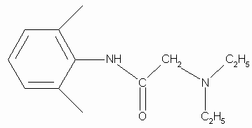
LIDOCAINE


2-(Diethylamino)-N-(2,6-dimethylphenyl)acetamide
Lidocaine is used in drugs formulated for local anaesthetics.
MW= 234.34 g/mol; MP= 68-69 o C; bruttoformula: C14H22N2O;
CAS-number: 137-58-6;

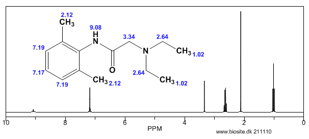
Lidocaine is an antiarrhythmic medicine and also serves as a local anaesthetic drug. It is utilized in topical application to relieve pain, burning and itching sensation caused from skin inflammations. This drug is mainly used for minor surgeries. Figure 1 shows the 1H NMR spectrum of 200 mM lidocaine in CDCl3.
Figure 1. Proton NMR spectrum of 200 mM lidocaine in CDCl3.
Calculated H-NMR spectrum of lidocaine

1H NMR Relaxation

Figures 2, 3 and 4 show the relaxation time measurements. It can be seen that the relaxation times are shortest for the CH2 protons and longest for the CH protons. The first data point amplitude increases with the number of protons for the related peak.
Figure 2. Proton T1 relaxation time measurement of 200 mM lidocaine in CDCl3.
Figure 3. Proton T2 relaxation time measurement of 200 mM lidocaine in CDCl3.
Figure 4. COSY spectrum of 200 mM lidocaine in CDCl3. The cross-peaks and corresponding exchanging protons are labeled by colour-coded arrows and ellipses.

2D COSY

Figure 4 shows the 2D COSY spectrum where two spin systems (6,7,8) to (10,11) can be clearly seen. For instance, the methyl groups at 10 and 11 positions bond to aromatic protons at 6 and 8 positions, while the methyl groups at 16 and 17 positions bond to the ethylene groups at 14 and 15 positions. No coupling occurs at positions (6,7,8) to (16,17) or (14,15).

2D Homonuclear J-Resolved Spectroscopy

The chemical shift in the 2D homonuclear j-resolved spectrum appears along the direct (f2) direction and the effects of coupling between protons appear along the indirect (f1) dimension. This enables the assignment of chemical shifts of multiplets and may help in measuring unresolved couplings. Also, a decoupled 1D proton spectrum is produced by the projection along the f1 dimension. The 2D homonuclear j-resolved spectrum of lidocaine, plus the 1D proton spectrum (blue line) are shown in Figure 5.
Figure 5. Homonuclear j-resolved spectrum of 200 mM lidocaine in CDCl3. The multiplet splitting frequencies for different couplings are colour- coded.
The projection which is vertical reveals how the multiplets disintegrate into a single peak, which makes the 1D spectrum more simplified. Peak multiplicities are produced by vertical traces from peaks in the 2D spectrum and help in determining the frequencies of proton-proton coupling. When coupling frequencies are compared between different peaks, information can be obtained regarding which peaks are bonded to each other. Also, Information regarding the coupling strength can be obtained from the size of the coupling frequencies. These couplings substantiate the results of the COSY experiment.
However, in this experiment, the effects of second order coupling appear in the f1 direction as additional peaks which are equidistant from the coupling partners detached from the zero frequency in the f1 dimension. These peaks provide proof of second order coupling partners, but are generally considered as artifacts. Figure 6 shows these coupling partners and additional peaks marked by colour-coded arrows and ellipses.
Figure 6. Homonuclear j-resolved spectrum of 200 mM lidocaine in CDCl3 showing the extra peaks due to strong couplings.

1D 13C Spectra

Figure 7 shows the 13C NMR spectra of 1 M lidocaine in CDCl3. Since the 1D Carbonexperiment is highly susceptible to the 13C nuclei in the specimen, it easily and clearly resolves 9 resonances. In this experiment, only carbons coupled to protons are seen.
Figure 7. Carbon spectra of 1 M lidocaine in CDCl3.
Given the fact that the DEPT spectra do not display the peaks at 170 and 135ppm, they must be part of quaternary carbons. The DEPT-135 and the DEPT-45 experiments provide signals ofCH3, CH2 and CH groups, while the DEPT-90 experiment provides only the signal of CH groups. However, in DEPT-135 the CH2 groups occur as negative peaks. It can thus be summed up that the peaks between 45 and 60ppm belong to ethylene groups; the peaks between 10 and 20ppm are part of the methyl groups; and the peaks between 125 and 130ppm belong to methyne groups. A similar study can be carried out on the C and CH peaks.

Heteronuclear Correlation

The Heteronuclear Correlation (HETCOR) experiment identifies the proton signal that appears along the indirect dimension and the carbon signal along the direct dimension. Figure 8 shows the HETCOR spectrum of 1 M lidocaine in CDCl3. in the 2D spectrum, the peaks reveal which proton is attached to which carbon. This experiment helps in resolving assignment uncertainty from the ID carbon spectra.
Figure 8. HETCOR spectrum of 1 M lidocaine in CDCl3.

Heteronuclear Multiple Quantum Coherence

Heteronuclear Multiple Quantum Coherence (HMQC) is similar to the HETCOR experiment and is utilized to associate proton resonances to the carbons that are coupled directly to those protons. But in the HMQC experiment, the proton signal appears along the direct dimension and the carbon signal along the indirect dimension. Figure 9 shows the HMQC spectrum of 1 Mlidocaine in CDCl3. In the 2D spectrum, the peaks show which proton is attached to which carbon. For conclusive peak assignment, a similar study with the HETCOR spectrum can be carried out.
Figure 9. HMQC spectrum of 1 M lidocaine in CDCl3.

Heteronuclear Multiple Bond Correlation

The Heteronuclear Multiple Bond Correlation (HMBC) experiment can be employed to achieve long-range correlations of proton and carbon via two or three bond couplings. Similar to the HMQC experiment, the proton signal appears along the direct dimension and the carbon signal along the indirect dimension. Figure 10 shows the HMBC spectrum of 1 M lidocaine in CDCl3.
Figure 10. HMBC spectrum of 1 M lidocaine in CDCl3, with some of the long-range couplings marked.
The couplings amid the molecular positions appear analogous to the couplings seen in the COSY spectrum; however, the HMBC also displays couplings to quaternary carbons, which are not seen either in HMQC or COSY experiments. In addition, there is a correlation between protons and carbons. This is attributed to three-bond bonding from 14 and 15 and vice versa, as shown in light green in Figure 1.
IR

Chemical reaction of lidocaine with singlet oxygen. Rate constants for the chemical reaction between lidocaine and O2(1D g) were determined in methanol, acetonitrile and N,N-dimethylformamide. Lidocaine consumption was followed during the reaction. Rate constants for the chemical reaction, kRLID, are (1.05 ± 0.061) x 10M-1 s-1, (1.42 ± 0.073) x 10M-1 s-1and (0.61 ± 0.046) x 10M-1 s-1 in acetonitrile, methanol and N,N-dimethylformamide, respectively.
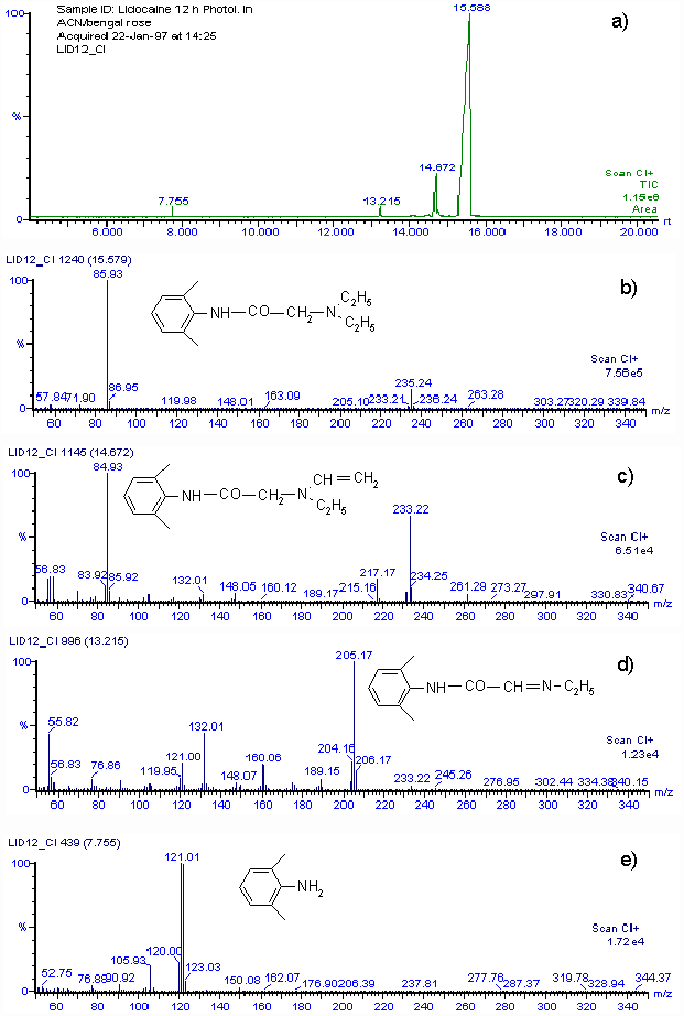
By using the Mair method (4) for hydroperoxide determination, a concentration equivalent to 0.0153 M of hydroperoxide was found when 0.03 M lidocaine in acetonitrile was irradiated for 12 h in the presence of Rose Bengal. The amount of hydroperoxide produced agrees with the consumption of lidocaine. Although we cannot isolate reaction products in quantities adequate for spectroscopic characterization, a rough idea of the product distribution was obtained by GC-MS analysis of the main lidocaine derivatives produced in the photooxidations. When 0.03 M lidocaine was irradiated for 12 h in the presence of Rose Bengal the results shown in Fig. 2 a) are obtained with the mass spectrometer in the positive chemical ionization (CI+) mode. Only four peaks appear in the chromatogram, the main one, with a retention time of 15.59 min, is that of unreacted lidocaine. Fig. 2 b) shows that the mass spectrum is that of lidocaine. The CI+ and EI (not included) mass spectra corresponding to peaks at retention times of 14.57, 13.22 and 7.77 min, indicate that 2-(ethylvinylamino)-N-(2,6-dimethylphenyl)-acetamide, 2-(1-azapropily-den)-N-(2,6-dimethyl-phenyl)-acetamide and 2,6-dimethylaniline are the probable main products of photooxidation of lidocaine. Figs. 2 c), 2 d) and 2 e), show the CI+ mass spectra and corresponding structures.

Figure 2.a) GC-MS chromatogram of 30 mM lidocaine in acetonitrile after 12 h of irradiation in the presence of Rose Bengal. b) CI+ mass spectrum of compound with retention time 15.58 m. c) CI+ mass spectrum of compound with retention time 14.67 m. d) CI+ mass spectrum of compound with retention time 13.22 m. e) CI+ mass spectrum of compound with retention time 7.76 m.