DR ANTHONY MELVIN CRASTO,WorldDrugTracker, helping millions, A 90 % paralysed man in action for you, I am suffering from transverse mylitis and bound to a wheel chair, With death on the horizon, nothing will not stop me except God................DR ANTHONY MELVIN CRASTO Ph.D ( ICT, Mumbai) , INDIA 25Yrs Exp. in the feld of Organic Chemistry,Working for GLENMARK GENERICS at Navi Mumbai, INDIA. Serving chemists around the world. Helping them with websites on Chemistry.Million hits on google, world acclamation from industry, academia, drug authorities for websites, blogs and educational contribution

Saturday, 2 January 2016

A dihydropyrrolizinyl oxindole


NOESY


General procedure for synthesis of compounds 7a–7c from the three-component reaction of isatins, proline and aroylacrylic acids: A mixture of isatin (1.0 mmol), proline (1.0 mmol) and aroylacrylic acid (1.0 mmol) in 4.0 mL aqueous ethanol (1:3) was heated in an oil bath to reflux temperature for 15 min. The resulting precipitates were collected by filtration and washed with cold ethanol to give analytically pure products 7. 7a: orange powder, 22%, mp 215–216 °C. 1H (500 MHz, DMSO-d6) and 13C NMR (125 MHz, DMSO-d6) data are given in Table 3. MS (m/z) (%): 462 (M+, 52), 435 (45), 405 (8), 353 (12), 317 (18), 289 (52), 208 (30), 173 (33), 127 (46), 75 (30), 41 (100); anal. calcd for C21H15BrСl2N2O (462.17): C 54.57; H 3.27; N 6.06; found: C 54.48; H 3.19; N 6.10.


The tentative mechanism for the formation of 7a is outlined in Scheme 3. First, the initially formed spiropyrrolizidine undergoes decarboxylation via ring opening of the spiro cycle. The subsequent enolization of the intermediate leads to the formation of the dihydropyrrolizinyl oxindole system.
[1860-5397-10-8-i3]
Scheme 3: Tentative reaction mechanism for the decarboxylative cyclative rearrangement of the initial three-component product.


For assigning structures of byproducts we carried out the reaction of isatins 1, aroylacrylic acids 5 and proline in a boiling mixture of EtOH and water, which resulted in the formation and isolation of compounds 7a–7c (Scheme 2). The unexpected structure of rearranged product 7a was confirmed by 1H, 13C and 2D NMR spectroscopy (Table 3).
[1860-5397-10-8-i2]
Scheme 2: The synthesis of compounds 7a–7c.

Table 3: 13C and 1H spectral data for compound 7a.
entryfunctional group13C1H
δ, ppmδ, ppmmultiplicityJ, Hz

11-NH10.59s
22-CO178.09
33-CH45.334.56s
43a-C (oxindole)134.06
54-CH (oxindole)127.557.05s
65-C (oxindole)113.56
76-CH (oxindole)130.777.33dd8.1; 2.2
87-CH (oxindole)111.576.80d8.1
97a-C (oxindole)142.21
105-C124.67
116-C120.38
127-CH99.695.30s
137a-C138.34
141-CH224.432.85–2.63m
152-CH227.312.43–2.27m
163-CH246.264.20–4.02, 3.90–3.70m
171-C Ar129.50
182-CH Ar129.877.82d1.8
193-C Ar133.12
204-C Ar131.63
215-CH Ar130.957.65d8.1
226-CH Ar128.447.55dd8.2; 1.8
The main feature of the 13C spectra of compounds 7a–7c is the absence of the signal of the 3C-spiro nucleus. The 1H NMR spectrum of compound 7a displays a singlet at 5.30 ppm for the 7-CH of the dihydropyrrolizinyl moiety, which shows a H,H-NOESY correlation with a singlet at 4.56 ppm (3-CH of the oxindole ring) and HMBCs with 7a-C at 138.34 ppm. The singlet at 4.56 ppm of 3-CH of the oxindole ring shows H,H-COSY and H,H-NOESY correlations with a singlet at 7.05 ppm of 4-CH (oxindole ring) and HMBCs with 2-CO at 178.09 ppm, 4-C at 127.55 ppm and 6-C at 120.38 ppm (Figure 7). The NH proton of the oxindole ring gives a singlet at 10.59 ppm.
[1860-5397-10-8-7]
Figure 7: The selected COSY, NOESY and HMBC correlations of the signals in the 1H and 13C NMR spectra of compound 7a.


see................http://www.beilstein-journals.org/bjoc/single/articleFullText.htm?publicId=1860-5397-10-8

The regioselective synthesis of spirooxindolo pyrrolidines and pyrrolizidines via three-component reactions of acrylamides and aroylacrylic acids with isatins and α-amino acids

Tatyana L. Pavlovskaya1, Fedor G. Yaremenko1,2, Victoria V. Lipson1,2,3Email of corresponding author, Svetlana V. Shishkina1, Oleg V. Shishkin1,3, Vladimir I. Musatov1 and Alexander S. Karpenko4
1State Scientific Institution “Institute for Single Crystals” of National Academy of Sciences of Ukraine, 60, Lenin ave., Kharkov, 61178, Ukraine
2Antidiabetic Drug Laboratory, State Institution “V.J. Danilevsky Institute of Problems of Endocrine Pathology at the Academy of Medical Sciences of Ukraine”, 10, Artem St., Kharkov, 61002, Ukraine
3Organic Chemistry Department, V.N. Karazin Kharkov National University, 4, Svobody Sq., 61077, Kharkov, Ukraine,
4A.V. Bogatsky physico-chemical institute of the National Academy of Sciences of Ukraine, 86, Lustdorfskaya doroga, 65080, Odessa, Ukraine
Email of corresponding author Corresponding author email     
This article is part of the Thematic Series "Multicomponent reactions II".
Guest Editor: T. J. J. Müller
Beilstein J. Org. Chem. 2014, 10, 117–126.


 GOA FOOD,  INDIA, GOA


 KING FISH PICKLE

Mojea Goa Amchi Mumbai


 Mojea Goa Amchi Mumbai


 Prawns Moile..... Sweet, Sour and Spicy......
 

 Goan Sausages Grays made with Onions and Potatoes n fried with a hint of Vinegar and Coconut Oil severed with Pav....

 Mojea Goa Amchi Mumbai


Yummy Juicy Mankios......
— with Francisco Xavier Dcosta Goa, Thebigcountryband Goa, Pudhari Goa and 19 others.
 
 
 Mojea Goa Amchi Mumbai


All set to cook green masala...
— with Colin Glen Fernandes, Sarita Uchil Shetty, Alcina Gomes and 15 others.

 
 Mojea Goa Amchi Mumbai

 Mojea Goa Amchi Mumbai


Posta Fish Rava Fry (King Fish)
— with Devyani Toraskar, Aah Goa, Fishermans Wharf Goa and 34 others.


GÖÄÑ FìSH CÜRRY RìCÉ

 DRY PRAWNS CURRY


PÖRK CHìLLY FRY. CÜT Ñ CLÉÄÑ PÖRK ìÑ TO MÉDìMìÜM ÖR SMÄLL PìÉCÉS, WÄSH Ñ MÄRìÑÄTÉ WìJH SÄLT ÖR VìÑÉGÄR FÖR MÄX 1 .HÖÜR THÉÑ ìÑ Ä PÅÑ ÖR KÄDÄìL ADD WÅTèR Ñ MÄRìÑATÉD MÉÄT Ñ
LÉT ìT ßÖìL ÖÑCÉ DÖNÉ ÄDD ÖìL Ñ FRY ìT ADD ßìÑDÄ SÖL (KÖKÜM FÜLL) ÖÑìÖÑS, GARLìC Ö GìÑGÉR FìÑÉ CHÖPPÉD, ÖÑìÖÑ, SALT Ñ SAFFRÖÑ POWDÉR Ä PìÑCH ÄÑD GRÉÉÑ CHìLLY SLìT FRY WÉLL Ñ RÉÄDY TÖ SÉRVè... TRY Ñ LÉT MÉ KÑÖW


/////////////

Zanthodione

.


Spectroscopic data
Zanthodione (I): orange needles; mp. 254-256 °C; 

UV (MeOH) λmax (log E) 239 (sh) (4.49), 249 (4.51), 392 (sh) (3.58), 408 (4.51), 428 (3.58) ñm; 

IR (KBr) vmax 3389 (OH); 1660, 1646 (C=0); 1601, 1460 (aromatic ring C=C); 1040, 94°6X(OCH20) cm-1

1H NMR (CDC13, 600 MHz) δ 6.40 (2H, s, OCH20), 7.73 (1H, br ddd, J = 8.4, 7.2, 1.8 Hz, H-10), 7.77 (1H, dt, J = 7.2, 1.2 Hz, H-9), 8.04 (1H, s, H-7), 8.26 (1H, s, NH exchangeable with D20), 8.29 (1H, dd, J = 7.2, 1.8 Hz, H-8), 9.26 (1H, d, J = 8.4 Hz, H-ll), 11.98 (1H, s, OH exchangeable with D20); 

EIMS m/z 307 [M]+ (90); 

HRESIMS m/z 307.1481 (caled for C17H9NOs, 307.1480).


Zanthodione (I) was obtained as orange needles. Its molecular formula C17H9NOs was established by EIMS ([M]+, m/z 307) and HRESIMS. The UV spectrum showed absorption máxima at 239 (sh), 240, 249, 392 (sh), 408, 428 nm, that indicated the presence of a 4,5-dioxoaporphine skeleton.11,12 Its IR spectrum indicated the presence of OH/NH (3000—3389 cm-1), two aryl ketone C=0 (1660, 1646 cm-1), one methylenedioxy (1040, 940 cm1) and a six-membered lactam (1587 cm-1) functionalities. The 1H NMR spectrum revealed five aromatic protons, two D20 exchangeable singlets of a H-bonded phenolic hydroxyl [8 11.98 (1H, s)], and an amidocarbonyl proton at 8 8.26 (1H, s), and a methylenedioxy group at 8 6.40 (2H, s). Five aromatic protons of the spectrum closely resembled those of other 7,8,9,10,11-unsubstituted dioxoaporphine.11,12 Therefore, the methylenedioxy group had to be on ring A. The singlet at δ 8.04 (1H, s) was ascribed to H-7 while H-8, H-9, H-10 and H-ll appeared as a complex ABCX coupling system. In agreement with the spectra of other C-11-unsubstituted aporphines, dehydro- and oxoaporphines, the most downfield doublet [δ 9.26 (1H, s)] was characteristic of H-ll, the 2H [δ 7.73 (1H, ddd, J = 8.4, 7.2, 1.8 Hz), 7.77 (1H, dt, J = 7.2, 1.2 Hz)] were assigned to H-10, 9, and another proton at 6 8.29 (1H, dd, J = 7.2, 1.8 Hz) was due to H-8. The correlations of H-7/NH, and H-8/H-7, 9 were also disclosed in the NOESY experiment (Fig. 1) and further supported the position of above assignments. By means of 1H-NMR, 1H,1H-COSY, and NOESY (Fig. 1) data, the structure of zanthodione was shownto be 4-hydroxy-7H-benzo[g][l,3]dioxolo[4',5';4,5 ]benzo[l,2,3-úfe]quinoline-5,6-dione (1).


 see...................
 http://www.scielo.cl/scielo.php?script=sci_arttext&pid=S0717-97072008000300019&lng=en&nrm=iso&ignore=.html


J. Chil. Chem. Soc, 53, N° 3 (2008) págs: 1631-1634

CHEMICAL CONSTITUENTS FROM THE STEM BARK OF ZANTHOXYLUM SCANDENS

MING-JEN CHENG1,2,3 CHUAN-FANG LIN1,3 HSUN-SHUO CHANG1, IH-SHENG CHEN1*
1Graduate Institute of Natural Products , College of Pharmacy, Kaohsiung Medical University, Kaohsiung, Taiwan 807, Republic of China.
2M.-J. C. and C.-F. L. contributed equally to the work in this paper.
Bioresource Collection and Research Center (BCRC), Food Industry Research and Development Institute (FIRDI), Hsinchu, Taiwan 300, Republic of China.


Journal of the Chilean Chemical Society

On-line version ISSN 0717-9707

J. Chil. Chem. Soc. vol.53 no.3 Concepción Sept. 2008

http://dx.doi.org/10.4067/S0717-97072008000300019 

////////////

Hyrtioerectine D, E F


Marinedrugs 11 01061 g001 1024


Hyrtioerectine D, E F


Figure 1. Structures of hyrtioerectines D–F (13) isolated from the marine sponge Hyrtios species.


Marinedrugs 11 01061 g002 1024


Figure 2. COSY and HMBC correlations observed for compound 1.

Structure Elucidation of Compounds 1–3

The HRMS of compound 1 established a molecular formula C20H13N3O4 requiring 16 degrees of unsaturation. Compound 1 gave a bluish color on the TLC with FeCl3 indicating its phenolic nature. Its 1H NMR spectrum (in methanol-d4, Table 1) showed resonances for eight aromatic protons ranging from 6.80 to 9.64 ppm, suggesting the absence of any aliphatic moiety in the molecule. The 13C NMR spectrum of 1 (Table 1) displayed resonances for 20 carbons including 8 methines and 12 quaternary carbons. Two of the 13C quaternary resonances were deshielded at δ 154.3 (C-6) and 153.0 (C-6′) suggesting the oxygenation of their corresponding carbons. Interpretation of the 1D (1H and 13C) and 2D (1H–1H COSY, HSQC and HMBC) NMR spectroscopic data supported the presence of two ABX systems for trisubstituted β-carboline (H-5, H-7 and H-8) and disubstituted indole (H-4ʹ, H-5ʹ and H-7ʹ) moieties, respectively. These two moieties include 15 degrees of unsatuartion suggesting the need for an additional degree of unsaturation in 1. Interpretation of the NMR data (1H, COSY and HSQC) supported the 3,4,6-trisubstitution of the β-carboline moiety. This was evident from the resonating signals at δ 8.07 (1H, d, J = 2.0, H-5), 6.80 (1H, dd, J = 8.5, 2.0, H-7), 7.30 (1H, d, J = 8.5, H-8) and 9.64 (s, H-1). Additional signals at δ 8.87 (1H, s, H-2ʹ), 7.57 (1H, d, J = 8.7, H-4ʹ), 7.13 (1H, dd, J = 8.7, 2.3, H-5ʹ) and 7.60 (1H, d, J = 2.3, H-7ʹ) were assigned as 3,6-disubstituted indole moiety. The HSQC experiment allowed the unambiguous assignment of the protonated methine carbons in 1 (Table 1). The quaternary carbons were unambiguously assigned from HMBC data (Table 2 and Figure 2). For example, the location of the OH moieties at C-6 and C-6′ was supported from HMBC correlations of H-5/C-6, H-7/C-6, H-8/C-6 and H-4′/C-6′, H-5′/C-6′ and H-7′/C-6′ (Table 2). The connection of the indole and β-carboline moieties through C-3 and C-3ʹ was supported from a 3JCH HMBC cross peak between H-2ʹ of the indole moiety (δ 8.87) and C-3 of the β-carboline moiety (δ 138.1). Finally, the quaternary carbon resonating at δ 173.0 (C-8′) was assigned as a carboxylic moiety at C-4 completing the degrees of unsaturation in 1. This was supported by an IR band at 1725 cm−1 for the carbonyl moiety of the carboxylic acid. Furthermore, the MS fragment ion at m/z 315.1009 with the molecular formula C19H13N3O2 [M − COOH + H]+ supported the presence of the carboxylic functionality, thus completing the assignment of 1.
Compound 2displayed a pseudomolecular ion peak at m/z 374.1138 corresponding to C21H16N3O4 [M + H]+, being 14 mass unit (CH2) more than 1 and requiring 16 degrees of unsaturation. Comparison of the NMR data of 2 with those of 1 showed identical similarity of the 1H and 13C NMR resonances of both compounds (Table 1). Moreover, a new three-proton singlet in 1H NMR at δ 3.84 (H3-9ʹ) correlates in the HSQC with the 13C NMR signal at δ 55.8 (C-9ʹ) suggesting the presence of a methoxyl moiety in 2 at C-6. The assignment of the methoxyl moiety at C-6 was secured from HMBC cross peak of OCH3/C-6 (Table 2). Additionally, the unambiguous assignments of the protonated and quaternary carbons in 2 were secured from HSQC and HMBC experiments, respectively, thus completing the assignment of 2.


Hyrtioerectine D (1): yellow solid; UV (MeOH) λmax (log ε) 387 nm (3.85), 248 nm (3.85), 312 nm (4.05); IR νmax (film) 3600, 3500, 3020, 2980, 1725, 1610, 1405, 1290, 1255, 1220, 890 cm−1; NMR data, see Table 1, Table 2; positive HRESIMS m/z 360.0981 (calcd for C20H14N3O4 [M + H]+, 360.0984).
Hyrtioerectine E (2): yellow solid; UV (MeOH) λmax (log ε) 420 nm (3.90), 252 nm (3.95), 315 nm (4.15); IR νmax (film) 3595, 3545, 3015, 2980, 1720, 1612, 1410, 1290, 1250, 1220, 895 cm−1; NMR data, see Table 1, Table 2; positive HRESIMS m/z 374.1138 (calcd for C21H16N3O4 [M + H]+, 374.1140).
Hyrtioerectine F (3): yellow solid; UV (MeOH) λmax (log ε) 380 nm (3.95), 240 nm (4.10), 315 nm (4.00); IR νmax (film) 3590, 3500, 3350, 3010, 2985, 1655, 1615, 1405, 1290, 1255, 1215, 895 cm−1; NMR data, see Table 1, Table 2; positive HRESIMS m/z 359.1142 (calcd for C20H15N4O3 [M + H]+, 359.1144).


Table 1. NMR spectroscopic data for hyrtioerectines D–F (13) (in methanol-d4).
Hyrtioerectine DHyrtioerectine EHyrtioerectine F
PositionδC (mult.) aδH, mult. (J in Hz)δC (mult.) aδH, mult. (J in Hz)δC (mult.) aδH, mult. (J in Hz)
1140.8 (CH)9.64, s141.2 (CH)9.66, s139.6 (CH)9.71, s
3138.1 (qC)138.2 (qC)138.3 (qC)
4138.8 (qC)138.8 (qC)138.8 (qC)
4a115.9 (qC)115.8 (qC)115.9 (qC)
4b130.1 (qC)130.3 (qC)130.4 (qC)
5108.2 (CH)8.07, d (2.0)110.1 (CH)8.10, d (2.2)107.2 (CH)8.09, d (2.2)
6154.3 (qC)156.2 (qC)154.2 (qC)
7113.5 (CH)6.80, dd (8.5, 2.0)113.5 (CH)6.78, dd (8.5, 2.2)113.7 (CH)6.76, dd (8.5, 2.2)
8113.1 (CH)7.30, d (8.5)113.1 (CH)7.27, d (8.5)113.5 (CH)7.32, d (8.5)
8a132.1 (qC)132.2 (qC)132.4 (qC)
9a133.2 (qC)133.1 (qC)133.2 (qC)
2′119.6 (CH)8.87, s119.5 (CH)8.88, s119.6 (CH)8.87, s
3′123.2 (qC)123.2 (qC)123.2 (qC)
3a′137.5 (qC)137.5 (qC)137.5 (qC)
4′114.1 (CH)7.57, d (8.7)114.1 (CH)7.61, d (8.7)114.1 (CH)7.64, d (8.0)
5′119.7 (CH)7.13, dd (8.7, 2.3)119.6 (CH)7.15, dd (8.7, 2.2)119.7 (CH)7.17, dd (8.7, 2.0)
6′153.0 (qC)153.1 (qC)153.0 (qC)
7′106.7 (CH)7.60, d (2.3)106.9(CH)7.62, d (2.2)106.7 (CH)7.65, d (2.0)
7a′132.6 (qC)132.5 (qC)132.6 (qC)
8′173.0 (qC)173.0 (qC)164.2 (qC)
9′55.8 (CH3)3.84, s
a multiplicities of the signals were obtained from HSQC experiments; qC = quaternary carbon, CH = methine; CH3 = methyl.

Table 2. Selected HMBC correlations for hyrtioerectines D–F (13) (in methanol-d4).
C#Hyrtiooerectine D (1)Hyrtioerectine E (2)Hyrtioerectine F (3)
HMBC (H→C#)HMBC (H→C#)HMBC (H→C#)
3H-1, H-2′H-1, H-2′H-1, H-2′
4---
4aH-1, H-5H-1H-1, H-5
4bH-8H-8H-8
5H-7H-7H-7
6H-5, H-7, H-8H-5, H-7, H-8, H3-9′H-5, H-8
7H-5H-5H-5
8H-7H-7H-7
8aH-5, H-7H-5, H-7H-5, H-7
9aH-1H-1H-1
2′---
3′H-2′H-2′H-2′
3a′H-2′, H-4′, H-5′, H-7′H-2′, H-4′, H-7′H-2′, H-4′, H-7′
4′H-5′H-5′H-5′
5′H-4′, H-7′H-4′, H-7′H-4′, H-7′
6′H-4′, H-5′, H-7′H-4′, H-5′, H-7′H-4′, H-5′, H-7′
7′H-5′H-5′H-5′
7a′H-2′, H-4′, H-7′H-2′, H-4′, H-7′H-2′, H-4′, H-7′

see
 http://www.mdpi.com/1660-3397/11/4/1061/htm


Mar. Drugs 2013, 11(4), 1061-1070; doi:10.3390/md11041061
Article
Bioactive Compounds from the Red Sea Marine Sponge Hyrtios Species
Diaa T. A. Youssef 1,*, Lamiaa A. Shaala 2 and Hani Z. Asfour 3
1
Department of Natural Products, Faculty of Pharmacy, King Abdulaziz University, Jeddah 21589, Kingdom of Saudi Arabia
2
Natural Products Unit, King Fahd Medical Research Center, King Abdulaziz University, Jeddah 21589, Kingdom of Saudi Arabia
3
Department of Medical Parasitology, Faculty of Medicine, Princess Al-Jawhara Center of Excellence in Research of Hereditary Disorders, King Abdulaziz University, Jeddah 21589, Kingdom of Saudi Arabia
*Author to whom correspondence should be addressed; Tel.: +966-548-535-344; Fax: +966-269-516-96.


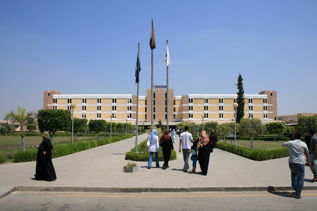
 Suez Canal University, Egypt


Map of suez canal university
Suez Canal University 
University in Ismaïlia, Egypt
The Suez Canal University is an Egyptian university serving the Suez Canal area, having its faculties divided among the Suez Canal governorates. It was established in 1964. It is notable for its non-classic research. Wikipedia
Address: EL SALAM DISTRICT، ISMAILEYA، Egypt


 ISMAILEYA، Egypt


//////////


////////////////////