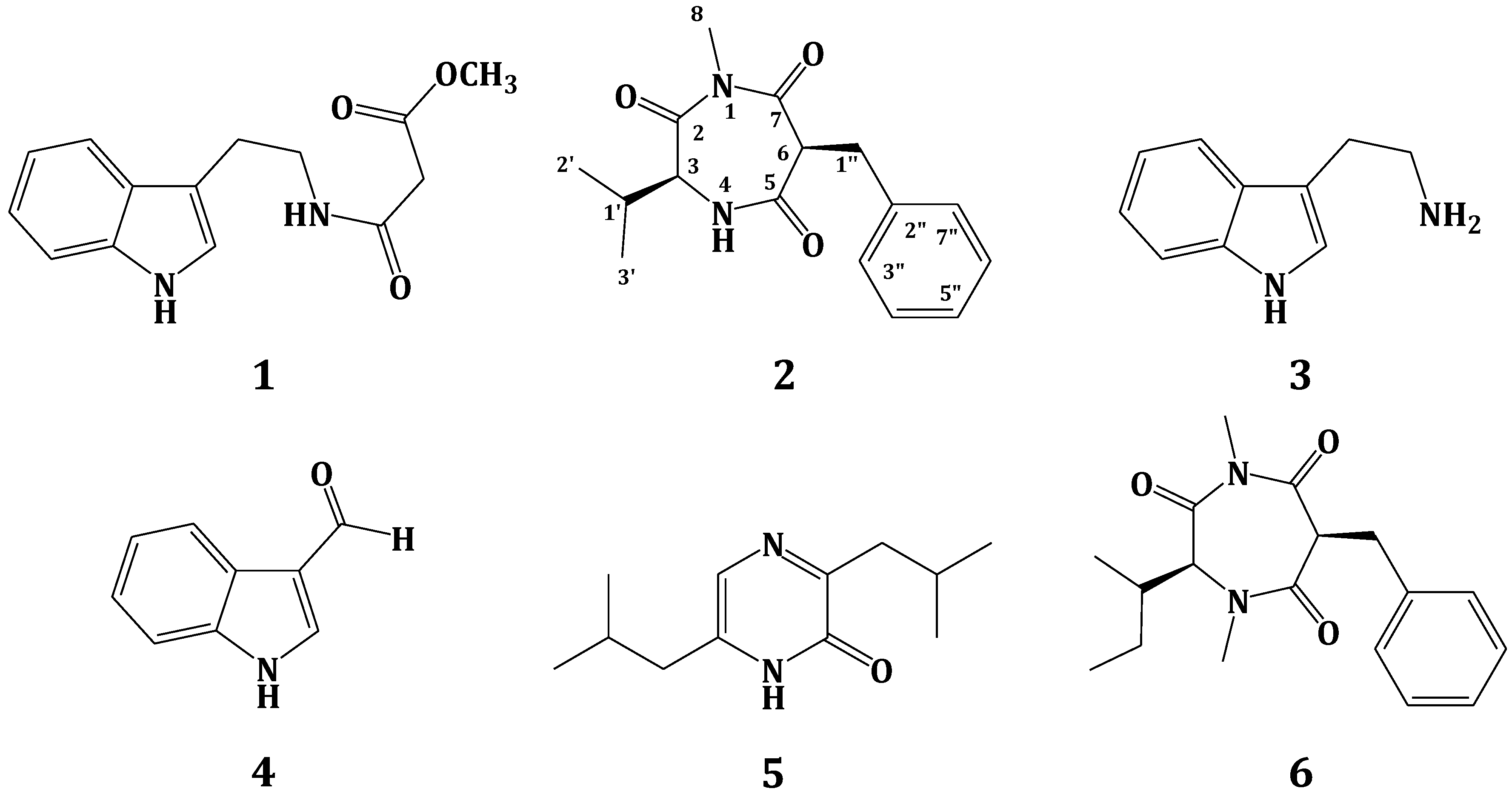
Hyrtioerectine D, E F
Figure 1. Structures of hyrtioerectines D–F (1–3) isolated from the marine sponge Hyrtios species.

Figure 2. COSY and HMBC correlations observed for compound 1.
Structure Elucidation of Compounds 1–3
The HRMS of compound 1 established a molecular formula C20H13N3O4 requiring 16 degrees of unsaturation. Compound 1 gave a bluish color on the TLC with FeCl3 indicating its phenolic nature. Its 1H NMR spectrum (in methanol-d4, Table 1) showed resonances for eight aromatic protons ranging from 6.80 to 9.64 ppm, suggesting the absence of any aliphatic moiety in the molecule. The 13C NMR spectrum of 1 (Table 1) displayed resonances for 20 carbons including 8 methines and 12 quaternary carbons. Two of the 13C quaternary resonances were deshielded at δ 154.3 (C-6) and 153.0 (C-6′) suggesting the oxygenation of their corresponding carbons. Interpretation of the 1D (1H and 13C) and 2D (1H–1H COSY, HSQC and HMBC) NMR spectroscopic data supported the presence of two ABX systems for trisubstituted β-carboline (H-5, H-7 and H-8) and disubstituted indole (H-4ʹ, H-5ʹ and H-7ʹ) moieties, respectively. These two moieties include 15 degrees of unsatuartion suggesting the need for an additional degree of unsaturation in 1. Interpretation of the NMR data (1H, COSY and HSQC) supported the 3,4,6-trisubstitution of the β-carboline moiety. This was evident from the resonating signals at δ 8.07 (1H, d, J = 2.0, H-5), 6.80 (1H, dd, J = 8.5, 2.0, H-7), 7.30 (1H, d, J = 8.5, H-8) and 9.64 (s, H-1). Additional signals at δ 8.87 (1H, s, H-2ʹ), 7.57 (1H, d, J = 8.7, H-4ʹ), 7.13 (1H, dd, J = 8.7, 2.3, H-5ʹ) and 7.60 (1H, d, J = 2.3, H-7ʹ) were assigned as 3,6-disubstituted indole moiety. The HSQC experiment allowed the unambiguous assignment of the protonated methine carbons in 1 (Table 1). The quaternary carbons were unambiguously assigned from HMBC data (Table 2 and Figure 2). For example, the location of the OH moieties at C-6 and C-6′ was supported from HMBC correlations of H-5/C-6, H-7/C-6, H-8/C-6 and H-4′/C-6′, H-5′/C-6′ and H-7′/C-6′ (Table 2). The connection of the indole and β-carboline moieties through C-3 and C-3ʹ was supported from a 3JCH HMBC cross peak between H-2ʹ of the indole moiety (δ 8.87) and C-3 of the β-carboline moiety (δ 138.1). Finally, the quaternary carbon resonating at δ 173.0 (C-8′) was assigned as a carboxylic moiety at C-4 completing the degrees of unsaturation in 1. This was supported by an IR band at 1725 cm−1 for the carbonyl moiety of the carboxylic acid. Furthermore, the MS fragment ion at m/z 315.1009 with the molecular formula C19H13N3O2 [M − COOH + H]+ supported the presence of the carboxylic functionality, thus completing the assignment of 1.Compound 2displayed a pseudomolecular ion peak at m/z 374.1138 corresponding to C21H16N3O4 [M + H]+, being 14 mass unit (CH2) more than 1 and requiring 16 degrees of unsaturation. Comparison of the NMR data of 2 with those of 1 showed identical similarity of the 1H and 13C NMR resonances of both compounds (Table 1). Moreover, a new three-proton singlet in 1H NMR at δ 3.84 (H3-9ʹ) correlates in the HSQC with the 13C NMR signal at δ 55.8 (C-9ʹ) suggesting the presence of a methoxyl moiety in 2 at C-6. The assignment of the methoxyl moiety at C-6 was secured from HMBC cross peak of OCH3/C-6 (Table 2). Additionally, the unambiguous assignments of the protonated and quaternary carbons in 2 were secured from HSQC and HMBC experiments, respectively, thus completing the assignment of 2.
Hyrtioerectine D (1): yellow solid; UV (MeOH) λmax (log ε) 387 nm (3.85), 248 nm (3.85), 312 nm (4.05); IR νmax (film) 3600, 3500, 3020, 2980, 1725, 1610, 1405, 1290, 1255, 1220, 890 cm−1; NMR data, see Table 1, Table 2; positive HRESIMS m/z 360.0981 (calcd for C20H14N3O4 [M + H]+, 360.0984).
Hyrtioerectine E (2): yellow solid; UV (MeOH) λmax (log ε) 420 nm (3.90), 252 nm (3.95), 315 nm (4.15); IR νmax (film) 3595, 3545, 3015, 2980, 1720, 1612, 1410, 1290, 1250, 1220, 895 cm−1; NMR data, see Table 1, Table 2; positive HRESIMS m/z 374.1138 (calcd for C21H16N3O4 [M + H]+, 374.1140).
Hyrtioerectine F (3): yellow solid; UV (MeOH) λmax (log ε) 380 nm (3.95), 240 nm (4.10), 315 nm (4.00); IR νmax (film) 3590, 3500, 3350, 3010, 2985, 1655, 1615, 1405, 1290, 1255, 1215, 895 cm−1; NMR data, see Table 1, Table 2; positive HRESIMS m/z 359.1142 (calcd for C20H15N4O3 [M + H]+, 359.1144).
| Hyrtioerectine D | Hyrtioerectine E | Hyrtioerectine F | ||||
|---|---|---|---|---|---|---|
| Position | δC (mult.) a | δH, mult. (J in Hz) | δC (mult.) a | δH, mult. (J in Hz) | δC (mult.) a | δH, mult. (J in Hz) |
| 1 | 140.8 (CH) | 9.64, s | 141.2 (CH) | 9.66, s | 139.6 (CH) | 9.71, s |
| 3 | 138.1 (qC) | 138.2 (qC) | 138.3 (qC) | |||
| 4 | 138.8 (qC) | 138.8 (qC) | 138.8 (qC) | |||
| 4a | 115.9 (qC) | 115.8 (qC) | 115.9 (qC) | |||
| 4b | 130.1 (qC) | 130.3 (qC) | 130.4 (qC) | |||
| 5 | 108.2 (CH) | 8.07, d (2.0) | 110.1 (CH) | 8.10, d (2.2) | 107.2 (CH) | 8.09, d (2.2) |
| 6 | 154.3 (qC) | 156.2 (qC) | 154.2 (qC) | |||
| 7 | 113.5 (CH) | 6.80, dd (8.5, 2.0) | 113.5 (CH) | 6.78, dd (8.5, 2.2) | 113.7 (CH) | 6.76, dd (8.5, 2.2) |
| 8 | 113.1 (CH) | 7.30, d (8.5) | 113.1 (CH) | 7.27, d (8.5) | 113.5 (CH) | 7.32, d (8.5) |
| 8a | 132.1 (qC) | 132.2 (qC) | 132.4 (qC) | |||
| 9a | 133.2 (qC) | 133.1 (qC) | 133.2 (qC) | |||
| 2′ | 119.6 (CH) | 8.87, s | 119.5 (CH) | 8.88, s | 119.6 (CH) | 8.87, s |
| 3′ | 123.2 (qC) | 123.2 (qC) | 123.2 (qC) | |||
| 3a′ | 137.5 (qC) | 137.5 (qC) | 137.5 (qC) | |||
| 4′ | 114.1 (CH) | 7.57, d (8.7) | 114.1 (CH) | 7.61, d (8.7) | 114.1 (CH) | 7.64, d (8.0) |
| 5′ | 119.7 (CH) | 7.13, dd (8.7, 2.3) | 119.6 (CH) | 7.15, dd (8.7, 2.2) | 119.7 (CH) | 7.17, dd (8.7, 2.0) |
| 6′ | 153.0 (qC) | 153.1 (qC) | 153.0 (qC) | |||
| 7′ | 106.7 (CH) | 7.60, d (2.3) | 106.9(CH) | 7.62, d (2.2) | 106.7 (CH) | 7.65, d (2.0) |
| 7a′ | 132.6 (qC) | 132.5 (qC) | 132.6 (qC) | |||
| 8′ | 173.0 (qC) | 173.0 (qC) | 164.2 (qC) | |||
| 9′ | 55.8 (CH3) | 3.84, s | ||||
a multiplicities of the signals were obtained from HSQC experiments; qC = quaternary carbon, CH = methine; CH3 = methyl.
| C# | Hyrtiooerectine D (1) | Hyrtioerectine E (2) | Hyrtioerectine F (3) |
|---|---|---|---|
| HMBC (H→C#) | HMBC (H→C#) | HMBC (H→C#) | |
| 3 | H-1, H-2′ | H-1, H-2′ | H-1, H-2′ |
| 4 | - | - | - |
| 4a | H-1, H-5 | H-1 | H-1, H-5 |
| 4b | H-8 | H-8 | H-8 |
| 5 | H-7 | H-7 | H-7 |
| 6 | H-5, H-7, H-8 | H-5, H-7, H-8, H3-9′ | H-5, H-8 |
| 7 | H-5 | H-5 | H-5 |
| 8 | H-7 | H-7 | H-7 |
| 8a | H-5, H-7 | H-5, H-7 | H-5, H-7 |
| 9a | H-1 | H-1 | H-1 |
| 2′ | - | - | - |
| 3′ | H-2′ | H-2′ | H-2′ |
| 3a′ | H-2′, H-4′, H-5′, H-7′ | H-2′, H-4′, H-7′ | H-2′, H-4′, H-7′ |
| 4′ | H-5′ | H-5′ | H-5′ |
| 5′ | H-4′, H-7′ | H-4′, H-7′ | H-4′, H-7′ |
| 6′ | H-4′, H-5′, H-7′ | H-4′, H-5′, H-7′ | H-4′, H-5′, H-7′ |
| 7′ | H-5′ | H-5′ | H-5′ |
| 7a′ | H-2′, H-4′, H-7′ | H-2′, H-4′, H-7′ | H-2′, H-4′, H-7′ |
see
http://www.mdpi.com/1660-3397/11/4/1061/htm
Mar. Drugs
2013,
11(4),
1061-1070;
doi:10.3390/md11041061
Article
Bioactive Compounds from the Red Sea Marine Sponge Hyrtios Species
1
Department of Natural Products, Faculty of Pharmacy, King Abdulaziz University, Jeddah 21589, Kingdom of Saudi Arabia
2
Natural Products Unit, King Fahd Medical Research Center, King Abdulaziz University, Jeddah 21589, Kingdom of Saudi Arabia
3
Department of Medical Parasitology,
Faculty of Medicine, Princess Al-Jawhara Center of Excellence in
Research of Hereditary Disorders, King Abdulaziz University, Jeddah
21589, Kingdom of Saudi Arabia
*Author to whom correspondence should be addressed; Tel.: +966-548-535-344; Fax: +966-269-516-96.
Suez Canal University, Egypt
Suez Canal University
University in Ismaïlia, Egypt
The
Suez Canal University is an Egyptian university serving the Suez Canal
area, having its faculties divided among the Suez Canal governorates. It
was established in 1964. It is notable for its non-classic research. Wikipedia
Address: EL SALAM DISTRICT، ISMAILEYA، Egypt


ISMAILEYA، Egypt


//////////
 .
.