.
.
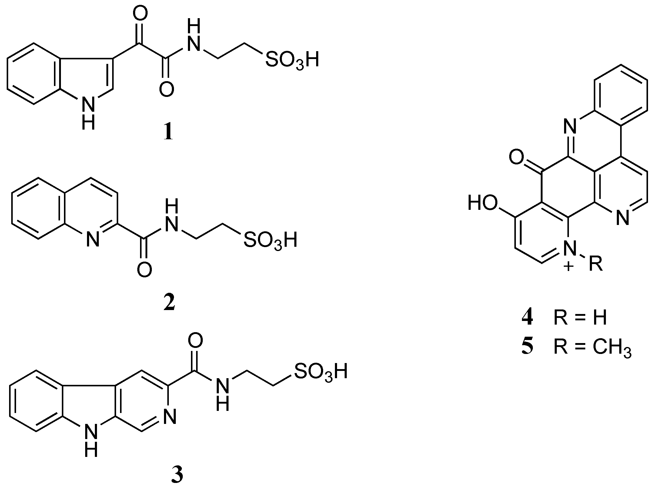
Figure 1. Structures of stolonines A-C (1–3), 11-hydroxyascididemin (4) and cnemidine A (5) isolated from the tunicate C. stolonifera.
| Position | δC | δH (J in Hz) | gCOSY | gHMBCAD |
|---|---|---|---|---|
| 1 | 12.20, br.s | 2 | ||
| 2 | 138.5 | 8.82, d (3.0) | 1 | 3, 3a, 7a |
| 3 | 112.1 | |||
| 3a | 126.2 | |||
| 4 | 121.3 | 8.22, dd (1.8, 7.2) | 5 | 6, 7a |
| 5 | 122.4 | 7.24, dt (1.8, 7.2) | 4, 6 | 3a, 7 |
| 6 | 123.3 | 7.26, dt (1.8, 7.2) | 5, 7 | 4, 7a |
| 7 | 112.5 | 7.52, dd (1.8, 7.2) | 6 | 3a, 5 |
| 7a | 136.2 | |||
| 8 | 181.6 | |||
| 9 | 162.7 | |||
| 10 | 8.78, t (5.4) | 11 | 9 | |
| 11 | 35.5 | 3.50, dd (6.0, 6.6) | 10, 12 | 9, 12 |
| 12 | 49.8 | 2.66, t (6.6) | 11 | 11 |
a 1H NMR at 600 MHz referenced to residual DMSO solvent (δH 2.50 ppm) and 13C NMR at 150 MHz referenced to residual DMSO solvent (δC 39.52 ppm).
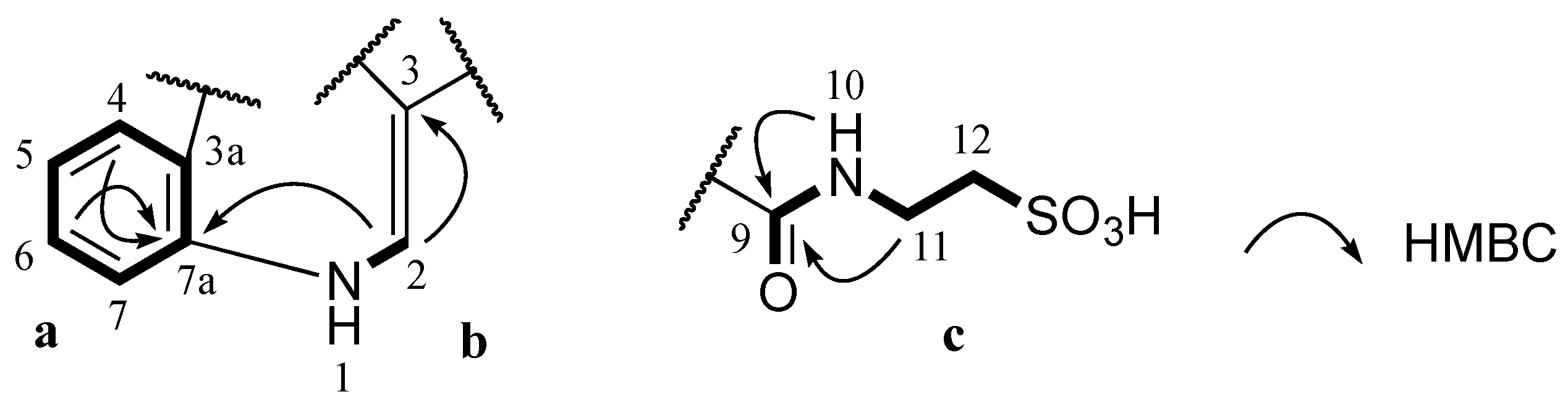
Stolonine A (1) was obtained as a white amorphous solid. The (−)-HRESIMS spectrum displayed a molecular ion [M−H]−at m/z 295.0390, which was consistent with the molecular formula C12H12N2O5S. The IR spectrum indicated the presence of an S=O stretching band at 1205 cm−1 [16]. A 1H NMR spectrum of 1 showed two exchangeable protons (δH 12.20 and 8.78 ppm), five aromatic protons (δH 8.82, 8.22, 7.52, 7.26 and 7.24 ppm) and two methylenes (δH 3.50 and 2.66 ppm). Further analysis of the 13C NMR and gHSQCAD spectra indicated that the molecule contained two carbonyls (δC 181.6 and 162.7 ppm), eight aromatic carbons (δC 138.5, 136.2, 126.2, 123.3, 122.4, 121.3, 112.5 and 112.1 ppm) and two methylenes (δC 49.8 and 35.5 ppm) (Table 1). J coupling constants of aromatic protons H-4 (δH 8.22, dd, 1.8, 7.2 Hz), H-5 (δH 7.24, dt, 1.8, 7.2 Hz), H-6 (δH 7.26, dt, 1.8, 7.2 Hz) and H-7 (δH 7.52, dd, 1.8, 7.2 Hz) and their COSY correlations were characteristic of a 1,2-disubstituted benzene ring (a, Figure 2). A gCOSY spectrum displayed correlations from the exchangeable proton H-1 (δH12.20, br.s) to H-2 (δH 8.82, d, 3.0 Hz) and also from H-11 (δH 3.50, dd, 6.0, 6.6 Hz) to the triplet exchangeable proton H-10 (δH 8.78, t, 5.4 Hz) and H-12 (δH 2.66, t, 6.6 Hz) facilitating the establishment of two other spin systems, –NH–CH= (b,Figure 2) and –NH–CH2–CH2– (c, Figure 2) respectively. The aromatic carbon C-3 (δC 112.1 ppm) was attached to C-2 (δC138.5 ppm) in the moiety b determined by a HMBC correlation from H-2 to C-3. A HMBC correlation from H-2 to C-7a (δC136.2 ppm) supported the linkage from a to b at C-7a (Figure 2). Both protons H-10 and H-11 in the moiety c showed HMBC correlations with a carbonyl carbon at δC 162.7 ppm suggesting the connection of this carbonyl to N-10 to form an amide bond (c, Figure 2). Two methylene signals at δH 3.50 and 2.66 ppm corresponding to carbons C-11 (δC 35.5 ppm) and C-12 (δC 49.8 ppm) as well as the relatively downfield resonance of the methylene C-12 were diagnostic of methylenes in a taurine moiety (c, Figure 2) [17,18,19].


Figure 2. Partial structures (a, b and c) of 1 and their key HMBC correlations.
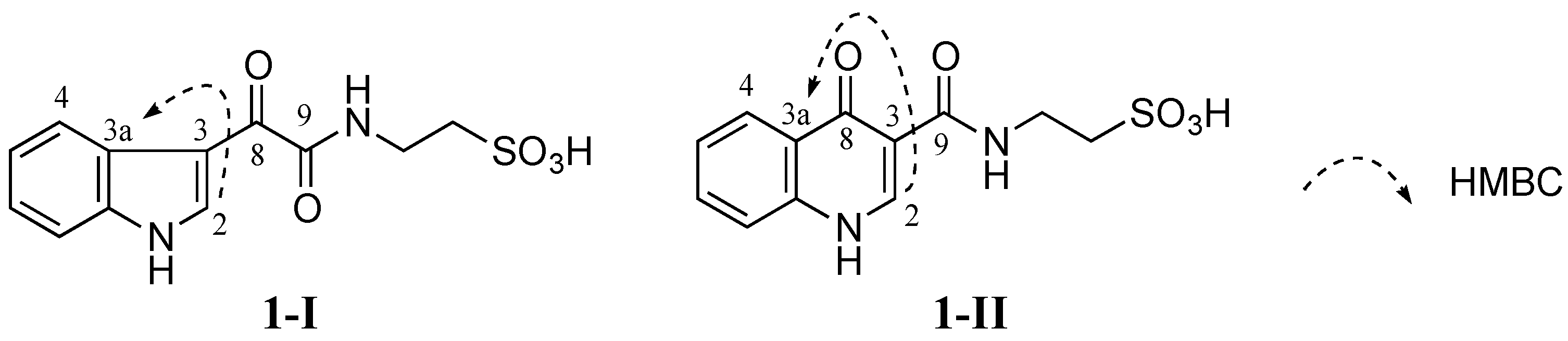
No HMBC correlation from any proton to the carbonyl C-8 (δC 181.6 ppm) was observed when HMBC experiments were performed and optimized with different JHC couplings. Therefore, two different isomers 1-I and 1-II were conceivable from these data (Figure 3). Detailed HMBC analysis showed that H-2 had a HMBC correlation with C-3a (δC 126.2 ppm). This suggested that 1 was favorable to 1-I since the H-2 to C-3a correlation in 1-I was a three-bond coupling while the H-2 to C-3a correlation in 1-II was a four-bond coupling (Figure 3).

Figure 3. Two possible structures 1-I and 1-II of 1 (1-I and 1-II are possible structural isomers of 1).
Figure 3. Two possible structures 1-I and 1-II of 1 (1-I and 1-II are possible structural isomers of 1).
| Position | δC (Exp.) | 1-I | 1-II | δH (Exp.) | 1-I | 1-II | ||||
|---|---|---|---|---|---|---|---|---|---|---|
| δC (Calc.) | δC (Scaled) | δC (Calc.) | δC (Scaled) | δH (Calc.) | δH (Scaled) | δH (Calc.) | δH (Scaled) | |||
| 2 | 138.5 | 136.9 | 140.3 | 141.3 | 142.8 | 8.82 | 8.74 | 8.61 | 8.84 | 8.67 |
| 3 | 112.1 | 111.5 | 112.9 | 111.2 | 110.2 | |||||
| 3a | 126.2 | 124.1 | 126.5 | 123.0 | 122.9 | |||||
| 4 | 121.3 | 118.9 | 120.9 | 124.3 | 124.4 | 8.22 | 8.62 | 8.48 | 8.40 | 8.19 |
| 5 | 122.4 | 120.1 | 122.1 | 121.1 | 120.9 | 7.24 | 7.63 | 7.40 | 7.63 | 7.35 |
| 6 | 123.3 | 120.9 | 123.0 | 129.4 | 129.9 | 7.26 | 7.59 | 7.36 | 8.00 | 7.76 |
| 7 | 112.5 | 108.7 | 109.9 | 114.8 | 114.1 | 7.52 | 7.55 | 7.31 | 7.53 | 7.24 |
| 7a | 136.2 | 131.9 | 134.9 | 134.4 | 135.3 | |||||
| 8 | 181.6 | 176.7 | 183.2 | 172.5 | 176.5 | |||||
| 9 | 162.7 | 156.8 | 161.7 | 159.4 | 162.4 | |||||
| 11 | 35.5 | 37.1 | 32.6 | 36.9 | 29.8 | 3.50 | 3.69 | 3.11 | 3.71 | 3.08 |
| 12 | 49.8 | 57.2 | 54.4 | 58.4 | 53.1 | 2.66 | 3.55 | 2.95 | 3.57 | 2.93 |
| CMAE | 1.5 | 3.1 | 0.23 | 0.25 | ||||||
| DP4 | 100.0% | 0.0% | 85.3% | 14.7% | ||||||
Stolonine A (1): white, amorphous solid; UV (MeOH) λmax (log ε) 210 (3.8), 252 (3.5) and 325 (3.3) nm; IR (film) νmax 3307, 1681, 1205, 1049 and 802 cm−1; 1H (600 MHz, DMSO-d6) and 13C (150 MHz, DMSO-d6) NMR data are summarized in Table 1; (−)-HRESIMS m/z 295.0390 [M − H]− (calcd for [C12H11N2O5S]−, 295.0394, Δ −1.4 ppm).
Mar. Drugs 2015, 13(7), 4556-4575; doi:10.3390/md13074556
Article
Isolation and Total Synthesis of Stolonines A–C, Unique Taurine Amides from the Australian Marine Tunicate Cnemidocarpa stolonifera
1
Eskitis Institute for Drug Discovery, Griffith University, Brisbane, Queensland 4111, Australia
2
Queensland Museum, Brisbane, Queensland 4101, Australia
*
Author to whom correspondence should be addressed; Tel.: +61-7-3735-6009; Fax: +61-7-3735-6001.
http://www.mdpi.com/1660-3397/13/7/4556/htm
/////////////////
RAIN FOREST, KODIAK ISLAND, ALASKA, USA


HI EVERYONE - YESTERDAY, WE WERE IN THE ROCKY MOUNTAINS.
TODAY, WE ARE GOING TO KODIAK ISLAND IN ALASKA
TO CHECK OUT A RAINFOREST.
IN ORDER TO BE CONSIDERED A RAINFOREST,
IT HAS TO RECEIVE BETWEEN 98 AND 180 INCHES OF
PERCIPITATION, INCLUDING SNOW. ALASKA'S RAINFOREST SANCTUARY IS A
TEMPERATE RAINFOREST, AS OPPOSED TO A TROPICAL RAINFOREST.
KODIAK ISLAND HAS FOUR SEASONS. SPRING HAS A
LOT OF RAIN, CAUSING TREES AND PLANTS TO BLOOM.
SUMMER HAS WARM TEMPERATURES OF 70 - 80 DEGREES FARENHEIT.
AUTUMN HAS COOL TEMPERATURES AND RAIN. WINTER HAS
VERY COLD TEMPERATURES AND SNOW. THE AVERAGE
ANNUAL TEMPERATURE IS 50 DEGREES F.
KODIAK ISLAND'S RAINFOREST RESERVE IS 40 ACRES AND
INCLUDES TALL STANDS OF SPRUCE, HEMLOCK AND
CEDAR TREES WITH A FOREST FLOOR SATURATED WITH MOSSES,
WILD FLOWERS AND A VARIETY OF BERRIES.
THE SANCTUARY IS LOCATED AT PISCURESQUE HERRING COVE.
ON-SITE EXPERIENCES INCLUDE INTERACTING WITH A
HERD OF ALASKAN REINDEER, VISITING THE ALASKA WILDLIFE
FOUNDATION CENTER, WATCHING A NATIVE MASTER TOTEM-POLE
CARVER AT WORK AND VISITING A HISTORIC ALASKAN SAWMILL.
THE ALASKA RAINFOREST SANCTUARY IS A UNIQUE ALASKA
EXPERIENCE AND PERFECT FOR THE NATURE LOVER.
EAGLE CREEK, ONE OF ALASKA'S RICHEST SALMON SPAWNING STREAMS,
FLOWS THROUGH THIS DIVERSE ECOSYSTEM, INTO THE OCEAN.
A MAJOR FISH HATCHERY IS LOCATED ACROSS
THE CREEK FROM THE SANCTUARY BOARDWALK.
I HOPE YOU HAVE ENJOYED YOUR VIST TO
KODIAK ISLAND, ALASKA TO SEE THE RAINFOREST.
RAINFOREST VEGETATION


BROWN BEAR


KODIAK BEAR FAMILY

LORIKEET BIRDS IN RAINFOREST - HARLEQUIN DUCK

BLUE GROUSE - CLARK'S NUTCRACKER
KODIAK ISLAND HAS LOTS OF BALD EAGLES

SWORD FERN - WILD STRAWBERRIES


HI EVERYONE - YESTERDAY, WE WERE IN THE ROCKY MOUNTAINS.
TODAY, WE ARE GOING TO KODIAK ISLAND IN ALASKA
TO CHECK OUT A RAINFOREST.
TODAY, WE ARE GOING TO KODIAK ISLAND IN ALASKA
TO CHECK OUT A RAINFOREST.
IN ORDER TO BE CONSIDERED A RAINFOREST,
IT HAS TO RECEIVE BETWEEN 98 AND 180 INCHES OF
PERCIPITATION, INCLUDING SNOW. ALASKA'S RAINFOREST SANCTUARY IS A
TEMPERATE RAINFOREST, AS OPPOSED TO A TROPICAL RAINFOREST.
IT HAS TO RECEIVE BETWEEN 98 AND 180 INCHES OF
PERCIPITATION, INCLUDING SNOW. ALASKA'S RAINFOREST SANCTUARY IS A
TEMPERATE RAINFOREST, AS OPPOSED TO A TROPICAL RAINFOREST.
KODIAK ISLAND HAS FOUR SEASONS. SPRING HAS A
LOT OF RAIN, CAUSING TREES AND PLANTS TO BLOOM.
SUMMER HAS WARM TEMPERATURES OF 70 - 80 DEGREES FARENHEIT.
AUTUMN HAS COOL TEMPERATURES AND RAIN. WINTER HAS
VERY COLD TEMPERATURES AND SNOW. THE AVERAGE
ANNUAL TEMPERATURE IS 50 DEGREES F.
LOT OF RAIN, CAUSING TREES AND PLANTS TO BLOOM.
SUMMER HAS WARM TEMPERATURES OF 70 - 80 DEGREES FARENHEIT.
AUTUMN HAS COOL TEMPERATURES AND RAIN. WINTER HAS
VERY COLD TEMPERATURES AND SNOW. THE AVERAGE
ANNUAL TEMPERATURE IS 50 DEGREES F.
KODIAK ISLAND'S RAINFOREST RESERVE IS 40 ACRES AND
INCLUDES TALL STANDS OF SPRUCE, HEMLOCK AND
CEDAR TREES WITH A FOREST FLOOR SATURATED WITH MOSSES,
WILD FLOWERS AND A VARIETY OF BERRIES.
THE SANCTUARY IS LOCATED AT PISCURESQUE HERRING COVE.
INCLUDES TALL STANDS OF SPRUCE, HEMLOCK AND
CEDAR TREES WITH A FOREST FLOOR SATURATED WITH MOSSES,
WILD FLOWERS AND A VARIETY OF BERRIES.
THE SANCTUARY IS LOCATED AT PISCURESQUE HERRING COVE.
ON-SITE EXPERIENCES INCLUDE INTERACTING WITH A
HERD OF ALASKAN REINDEER, VISITING THE ALASKA WILDLIFE
FOUNDATION CENTER, WATCHING A NATIVE MASTER TOTEM-POLE
CARVER AT WORK AND VISITING A HISTORIC ALASKAN SAWMILL.
THE ALASKA RAINFOREST SANCTUARY IS A UNIQUE ALASKA
EXPERIENCE AND PERFECT FOR THE NATURE LOVER.
EAGLE CREEK, ONE OF ALASKA'S RICHEST SALMON SPAWNING STREAMS,
FLOWS THROUGH THIS DIVERSE ECOSYSTEM, INTO THE OCEAN.
A MAJOR FISH HATCHERY IS LOCATED ACROSS
THE CREEK FROM THE SANCTUARY BOARDWALK.
I HOPE YOU HAVE ENJOYED YOUR VIST TO
KODIAK ISLAND, ALASKA TO SEE THE RAINFOREST.
KODIAK ISLAND, ALASKA TO SEE THE RAINFOREST.
RAINFOREST VEGETATION
BROWN BEAR
KODIAK BEAR FAMILY
LORIKEET BIRDS IN RAINFOREST - HARLEQUIN DUCK
BLUE GROUSE - CLARK'S NUTCRACKER
KODIAK ISLAND HAS LOTS OF BALD EAGLES
SWORD FERN - WILD STRAWBERRIES
Kodiak Island
Island in Alaska
Kodiak
Island is a large island on the south coast of the U.S. state of
Alaska, separated from the Alaska mainland by the Shelikof Strait. Wikipedia


Kodiak Municipal Airport, Kodiak Island, Alaska


////////////
 .
.


![2D [1H,1H]-TOCSY, 7.4 spectrum for Taurine](https://lh3.googleusercontent.com/blogger_img_proxy/AEn0k_u1k9tBzG0EnM0yd-j3hpIivU2N800ND6ZOQ0Hyy1qfbYEfZ9cMc-hVXC07ae5Zfq1OHgcWbIzoCDss6vOez-11-3VMbs5S7mBvkAwGJdF9KkHwNGBJ5qm0Awbm6iBPnAZEvEhTx7LYmeE4Gp2oOPE8xEI2cNpFA2BptwO9ipwbToIWjuCD=s0-d)


![2D [1H,13C]-HSQC, 7.4 spectrum for Taurine](https://lh3.googleusercontent.com/blogger_img_proxy/AEn0k_vjXyyJ6QxS9I3irUHjA_oMcilRUyrwB9neqQrebnwEk6tuXzZIbGakNLJ9fqNj68xwA7p8NtEm-iMPs0A1yzxlaT0BJ9jWl4TYW0ry80BvLh6yXx2aPrJf8uj_YmLp5kMmypFswPfGBIMpgui9gX7_jE9W1Mw9QGg83sXSFy3TZA3prn_V09Qj=s0-d)
![2D [1H,13C]-HMBC, 7.4 spectrum for Taurine](https://lh3.googleusercontent.com/blogger_img_proxy/AEn0k_ugBSceau1kkGRGevlJlYK0Vgl3OvIgJxr_8ttDHiRaK4H92L-zFk7mGjkusI3rLErfpxMMaoTzGtj4pW2UoHmXg55Sg3p1YmFQYwIeWdnDJqeJce1U1l408gqYQdjACQQyzztCMxFm7E2kBcFqbBHG3eMoAwXkz0RPGfmyXTuULRQVlZztOAQ=s0-d)