DR ANTHONY MELVIN CRASTO,WorldDrugTracker, helping millions, A 90 % paralysed man in action for you, I am suffering from transverse mylitis and bound to a wheel chair, With death on the horizon, nothing will not stop me except God................DR ANTHONY MELVIN CRASTO Ph.D ( ICT, Mumbai) , INDIA 25Yrs Exp. in the feld of Organic Chemistry,Working for GLENMARK GENERICS at Navi Mumbai, INDIA. Serving chemists around the world. Helping them with websites on Chemistry.Million hits on google, world acclamation from industry, academia, drug authorities for websites, blogs and educational contribution
Showing posts with label drugs. Show all posts

Saturday 17 October 2015

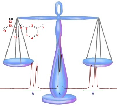
High-Throughput qNMR


It is well known that NMR is a very convenient technique for quantification, provided the amount of the material is within the limits of detection in NMR. 

When it comes to the actual calculations, this is a very straightforward process that does not require any fancy mathematics. Typically you will select the most convenient signal(s) or multiplet(s) in your spectrum and calculate the integral which can then be mapped to the corresponding concentration units by using a scaling factor that was previously calculated using some internal or external references.
read all at
http://nmr-analysis.blogspot.in/2012/12/high-throughput-qnmr.html

Infrared Spectroscopy,Characterisation of Organic Compounds

This discussion is on infrared (IR) spectroscopy which tells about the functional groups in the molecule.

William de Wiveleslie Abney

Almost any compound having covalent bonds absorbs various frequencies of electromagnetic radiation in the infrared region of the electromagnetic spectrum. This region lies at wavelength longer than those associated with visible light, which range from approximately 400 to 800 nm, but lies at wavelengths shorter than those associated with microwaves, which are longer than 1 mm. For chemical purposes, we are interested in the vibrational portion of the infrared region. It includes radiations with wavelengths between 2.5 μm and 25 μm.


In the early use of IR spectroscopy in 1882, William Abney managed to identify 52 benzene derivatives from the IR spectroscopy. IR spectroscopy is used to see the the functional group in the molecule as the bonds vibrates at certain wavenumber and it only vibrate at only certain allowable frequencies. 

The covalent bonds in a molecule can be described in similar way with a 2 balls (atoms) that connected with a spring (the bond). The bond distance continually changes, but an equilibrium or average bond distance can be defined. Whenever the spring is stretched or compressed beyond this equilibrium distance, the potential energy of system increases. As the spring moves in a harmonic oscillation, the energy is proportional to the frequency of vibration which is determined by the force constant (K) of the spring and the masses of two bonded atoms. The natural frequency of vibration of a bond is given by the equation
which is derived from Hooke's Law for vibrating springs. The reduced mass, μ, of the system is given by
From those equations, two things should be noticeable immediately. One is that stronger bonds have a larger force constant K and vibrate at higher frequencies than weaker bonds. Therefore, triple bonds will vibrate at higher frequencies (higher wavenumber) than double bonds or single bonds.
The second is that bonds between atoms of higher masses vibrate at lower frequencies than bonds between lighter atoms as shown below.
Besides that, when a bond vibrates, not all modes of vibration are allowed. When vibration modes do not provides no dipole moment change, so it is not allowed; Only vibration modes give dipole moment change that is allowed. The result of unallowed vibration modes is there is no peak or signal in the spectra.
1-octyne (left) and 4-octyne (right)


Alkenes

Alkenes show many more peaks than alkanes. The principal peaks of diagnostic value are the C-H stretching peaks for the spcarbon at value greater than 3000 cm-1, along with C-H peaks for the spcarbon atoms. Also the C=C stretching peak near 1650 cm-1, with higher intensity of cis- than trans- configuration.
cis-pentene (above) and trans-pentene (below)

O-H and N-H stretching

The signals for O-H and N-H stretching occur around 3300 cm-1, but they look different. O-H stretching broad bands centering between 3400 and 3300 cm-1. In solution, it will also be possible to observe a free O-H stretching band at about 3600 cm-1 (sharp and weaker) to the left of the hydrogen bonded O-H peak.
1-butanol

Meanwhile, primary amines, R-NH2, show two N-H stretching bands in the range 3500-3300 cm-1, whereas secondary amines, R2N-H, show only one band in that region. Tertiary amines will not show an N-H stretch. Because of these features, it is easy to differentiate among primary, secondary, and tertiary amines by inspection of the N-H stretch region.
butylamine (above), dibutylamine (middle), and tributylamine (below)

Carbonyl stretching

The carbonyl group is present in aldehydes, ketones, acids, esters, amides, acid chlorides, and anhydrides. This group absorbs strongly in the range from 1850 to 1650 cm-1 because of its large change in dipole moment. In figure below provides the normal bas value for the C=O stretching vibrations of the various functional groups. The C=O frequency of a ketone, which is approximately in the middle of the range, is usually considered the reference point for comparison of these values. In this section we will focus on aldehydes, ketones, acids, and esters.
Normal base value for the C=O stretching vibrations for carbonyl groups
Aldehydes show a very strong band for the carbony group that appear in the range of 1740-1725 cm-1. A very important doublet can be observed in the C-H stretch regio for the aldehyde C-H near 2850 and 2750 cm-1. The presence of this doublet allows aldehydes to be distinguished from other carbonyl-containing compounds. In the other sides, ketones show a very strong band for C=O group that appears in the range of 1720-1708 cm-1.
Nonanal (left) and 2-nonanone (right)

Carboxylic acids show a very strong band for the C=O group that appears in the range of 1730-1700 cm-1 and the O-H stretch appears in the spectrum as a very broad band extending from 3400 - 2400 cm-1. This broad band centers on about 3000 cm-1 and partially obsecures the C-H stretching bands. If the very broad O-H stretch band is seen, along with a C=O peak, it almost certainly indicates the compound is a carboxylic acid.
Nonanoic acid
Besides, there are variations of carbonyl compounds that can shift the C=O stretch frequency. The first one, there is conjugation of C=O with C=C, it lowers the stretching frequency to around 1680 cm-1.
4-methyl pent-3-en-2-one (mesityl oxide)
The carbonyl amide absorbs at an even lower frequency, 1640-1680 cm-1, but the carbonyl ester absorbs at higher frequency, 1730 - 1740 cm-1.
Butyl propanoate (left) and N-butyl propanamide (right)

Besides that, carbonyl groups in small rings (5 carbons or less) absorb at an even higher frequency.
The C=O stretching vibrations for cyclic ketones

Carbon-Nitrogen Stretching

The C-N stretching absorbs around 1200 cm-1, and as the bond stronger the C=N stretch absorbs around 1660 cm-1 and is much stronger than the C=C absorption in the same region. The nitriles group absorb strongly just above 2200 cm-1. The alkyne C=Csignal is much weaker and is just below 2200 cm-1.
Octanenitrile

To summarise this section, we will see the approximation range of those signals in IR spectroscopy.
IR spectroscopy correlation chart

Carbon-13 NMR


Carbon-13 NMR (13C NMR or sometimes simply referred to as carbon NMR) is the application of nuclear magnetic resonance (NMR) spectroscopy to carbon. It is analogous to proton NMR (1H NMR) and allows the identification of carbon atoms in an organic moleculejust as proton NMR identifies hydrogen atoms. As such 13C NMR is an important tool in chemical structure elucidation in organic chemistry. 13C NMR detects only the 13C isotope of carbon, whose natural abundance is only 1.1%, because the main carbon isotope,12C, is not detectable by NMR since it has zero net spin.


Implementation

13C NMR has a number of complications that are not encountered in proton NMR. 13C NMR is much less sensitive to carbon than 1H NMR is to hydrogen since the major isotope of carbon, the 12C isotope, has a spin quantum number of zero and so is not magnetically active and therefore not detectable by NMR. Only the much less common 13C isotope, present naturally at 1.1% natural abundance, is magnetically active with a spin quantum number of 1/2 (like 1H) and therefore detectable by NMR. Therefore, only the few 13C nuclei present resonate in the magnetic field, although this can be overcome by isotopic enrichment of e.g. protein samples. In addition, thegyromagnetic ratio (6.728284 107 rad T−1 s−1) is only 1/4 that of 1H, further reducing the sensitivity. The overall receptivity of 13C is about 4 orders of magnitude lower than 1H.

[1]
Another potential complication results from the presence of large one bond J-coupling constants between carbon and hydrogen (typically from 100 to 250 Hz). In order to suppress these couplings, which would otherwise complicate the spectra and further reduce sensitivity, carbon NMR spectra are proton decoupled to remove the signal splitting. Couplings between carbons can be ignored due to the low natural abundance of 13C. Hence in contrast to typical proton NMR spectra which show multiplets for each proton position, carbon NMR spectra show a single peak for each chemically non-equivalent carbon atom.
In further contrast to 1H NMR, the intensities of the signals are not normally proportional to the number of equivalent 13C atoms and are instead strongly dependent on the number of surrounding spins (typically 1H). Spectra can be made more quantitative if necessary by allowing sufficient time for the nuclei to relax between repeat scans.


High field magnets with internal bores capable of accepting larger sample tubes (typically 10 mm in diameter for 13C NMR versus 5 mm for 1H NMR), the use of relaxation reagents,[2] for example Cr(acac)3 (chromium (III) acetylacetonate, CAS number 21679-31-2), and appropriate pulse sequences have reduced the time needed to acquire quantitative spectra and have made quantitative carbon-13 NMR a commonly used technique in many industrial labs. Applications range from quantification of drug purity to determination of the composition of high molecular weight synthetic polymers.
13C chemical shifts follow the same principles as those of 1H, although the typical range of chemical shifts is much larger than for 1H (by a factor of about 20). The chemical shift reference standard for 13C is the carbons in tetramethylsilane (TMS), whose chemical shift is considered to be 0.0 ppm.
Typical chemical shifts in 13C-NMR

DEPT spectra


DEPT spectra of propyl benzoate
DEPT stands for Distortionless Enhancement by Polarization Transfer. It is a very useful method for determining the presence of primary, secondary andtertiary carbon atoms. The DEPT experiment differentiates between CH, CH2and CH3 groups by variation of the selection angle parameter (the tip angle of the final 1H pulse):
  • 135° angle gives all CH and CH3 in a phase opposite to CH2
  • 90° angle gives only CH groups, the others being suppressed
  • 45° angle gives all carbons with attached protons (regardless of number) in phase

Signals from quaternary carbons and other carbons with no attached protons are always absent (due to the lack of attached protons).
The polarization transfer from 1H to 13C has the secondary advantage of increasing the sensitivity over the normal 13C spectrum (which has a modest enhancement from the NOE (Nuclear Overhauser Effect) due to the 1H decoupling).

APT spectra 

Another useful way of determining how many protons a carbon in a molecule is bonded to is to use an Attached Proton Test, which distinguishes between carbon atoms with even or odd number of attached hydrogens. A proper spin-echo sequence is able to distinguish between S, I2S and I1S, I3S spin systems: the first will appear as positive peaks in the spectrum, while the latter as negative peaks (pointing downwards), while retaining relative simplicity in the spectrum since it is still broadband proton decoupled.
Even though this technique does not distinguish fully between CHn groups, it is so easy and reliable that it is frequently employed as a first attempt to assign peaks in the spectrum and elucidate the structure.[3]

  1. ^ R. M. Silverstein, G. C. Bassler and T. C. Morrill (1991). Spectrometric Identification of Organic Compounds. Wiley.
  2. ^ Caytan, Elsa; Remaud, Gerald S.; Tenailleau, Eve; Akoka, Serge, GS; Tenailleau, E; Akoka, S (2007). "Precise and accurate quantitative 13C NMR with reduced experimental time". Talanta 71 (3): 1016–1021. doi:10.1016/j.talanta.2006.05.075. PMID 19071407
  3. ^ Keeler, James (2010). Understanding NMR Spectroscopy (2nd ed.). John Wiley & Sons. p. 457. ISBN 978-0-470-74608-0.


Wednesday 17 June 2015

IR SPECTRUM OF IMPORTANT DRUGS


    BRIEF DESCRIPTION OF THE DRAWINGS


    • Figure 1B: Infrared spectrum of anhydrous Mycophenolate Mofetil hydrochloride.


    • Figure 2B: Infrared spectrum of Venlafaxine hydrochloride form I.


    • Figure 3B: Infrared spectrum of Venlafaxine hydrochloride form II.
     

    • Figure 4B: Infrared spectrum of Sertraline hydrochloride form II.


    • Figure 5B: Infrared spectrum of Sertraline hydrochloride form I.
     

    • Figure 6B: Infrared spectrum of Donepezil hydrochloride form II.

     
    • Figure 7B: Infrared spectrum of Donepezil hydrochloride form III.
     

    • Figure 8B: Infrared spectrum of Terbinafine hydrochloride form.
     

    • Figure 9B: Infrared spectrum of Cinacalcet hydrochloride.

     
    • Figure 10B: Infrared spectrum of Citalopram hydrobromide.

     
    • Figure 11 B: Infrared spectrum of Aripiprazole hydrochloride form A.
     

    • Figure 12B: Infrared spectrum of Pramipexole Monohydrochloride.

     
    • Figure 13B: Infrared spectrum of Moxifloxacin hydrochloride methylene dichloride solvate.
     

    • Figure 14B: Infrared spectrum of anhydrous Moxifloxacin hydrochloride form IV.


    • Figure 15B: Infrared spectrum of Moxifloxacin hydrochloride acetic acid solvate.

     
    • Figure 16B: Infrared spectrum of Moxifloxacin hydrochloride nitromethane solvate.


    • Figure 17B: Infrared spectrum of Duloxetine hydrochloride.


    • Figure 18B: Infrared spectrum of Linezolid hydrochloride.

     
    • Figure 19B: Infrared spectrum of Memantine hydrochloride.


    • Figure 20B: Infrared spectrum of Rimonabant Hydrochloride form I.

     
    • Figure 21 B: Infrared spectrum of Clopidogrel Hydrochloride form I.


    • Figure 22B: Infrared spectrum of Clopidogrel Hydrobromide form A.


    • Figure 23B: Infrared spectrum of Prasugrel Hydrochloride form B.
     

    • Figure 24B: Infrared spectrum of Prasugrel Hydrochloride acetonitrile solvate.

     
    • Figure 25B: Infrared spectrum of Raloxifene Hydrochloride form A.
     
    • .
    • Figure 26B: Infrared spectrum of Raloxifene Hydrochloride tetrahydrofurane solvate.
    •  
    •  
    •  
    • Figure 27B: Infr 
    • ared spectrum of Olanzapine Dihydrochloride form I.


    • Figure 28B: Infrared spectrum of Darifenacin Hydrobromide.
    • Figure 29B: Infrared spectrum of Sitagliptine Hydrochloride in amorphous form.
     
    • Figure 30B: Infrared spectrum of Vardenafil Dihydrochloride.


    • Figure 31 B: Infrared spectrum of Erlotinib Hydrochloride form A.


.


Deserts


The Karakum Desert, also spelled Kara Kum and Gara Gum is a desert  in Central Asia. It occupies about 70 percent, or 350,000 km², of the area of Turkmenistan. Covering much of present day Turkmenistan, the Karakum Desert lies east of the Caspian Sea, with the Aral Sea to the north and the Amu Darya river and the Kyzyl Kum desert to the northeast. In modern times, with the shrinking of the Aral Sea, the extended “Aral Karakum” has appeared on the former seabed, with an estimated area of 15,440 sq. The sands of the Aral Karakum are made up of a salt-marsh consisting of finely-dispersed evaporites and remnants of alkaline mineral deposits, washed into the basin from irrigated fields. The dusts blown on a powerful east-west airstream carry pesticide residues that have been found in the blood of penguins in Antarctica.10. Kara-Kum Desert, Uzbekistan / Turkmenistan


Lets read about top ten world’s largest deserts.


9. Great Sandy Desert, Australia

The Great Sandy Desert is a 360,000 km2 (140,000 sq mi) expanse in northwestern Australia. Roughly the same size as Japan, it forms part of a larger desert area known as the Western Desert. The vast region of Western Australia is sparsely populated, without significant settlements. The Great Sandy Desert is a flat area between the rocky ranges of the Pilbara  and the Kimberley. To the southeast is the Gibson Desert and to the east is the Tanami Desert. The Rudall River National Park and Lake Dora are located in the southwest while Lake Mackay is located in the southeast.


8. Chihuahuan Desert, Mexico

The Chihuahuan Desert is a desert that straddles the U.S.-Mexico border in the central and northern portions of the Mexican Plateau, bordered on the west by the extensive Sierra Madre Occidental range, and overlaying northern portions of the east range, the Sierra Madre Oriental. On the U.S.  side it occupies the valleys and basins of central and southern New Mexico, Texas  west of the Pecos River and southeastern Arizona; south of the border, it covers the northern half of the Mexican state of Chihuahua, most of Coahuila, north-east portion of Durango, extreme northern portion of Zacatecas  and small western portions of Nuevo León. It has an area of about 140,000 square miles. It is the third largest desert of the Western Hemisphere and is second largest in North America, after the Great Basin Desert.


7. Great Basin Desert, USA

The Great Basin is the largest watershed of North America which does not drain to an ocean. Water within the Great Basin evaporates since outward flow is blocked. The basin extends into Mexico and covers most of Nevada and over half of Utah, as well as parts of California, Idaho, Oregon and Wyoming. The majority of the watershed is in the North American Desert ecoregion, but includes areas of the Forested Mountain and Mediterranean California ecoregions. The Great Basin includes several metropolitan areas and Shoshone  Great Basin tribes. A wide variety of animals can be found in great basin desert. Look to the rocky slopes around the desert mountain ranges, you may spot a very rare desert bighorn sheep. Other mammals of the desert include kit fox, coyote, skunk, black-tailed jackrabbit, ground squirrels, kangaroo rat and many species of mice. Bird species are very diverse in desert oases.


6. Great Victoria Desert, Australia

The Great Victoria Desert is a barren, arid, and sparsely populated desert  ecoregion  in southern Australia. It falls inside the states of South Australia and Western Australia and consists of many small sandhills, grasslands  and salt lakes. It is over 700 kilometres (430 mi) wide (from west to east) and covers an area of 424,400 square kilometres (163,900 sq mi). The Western Australia Mallee shrub ecoregion lies to the west, the Little Sandy Desert to the northwest, the Gibson Desert and the Central Ranges xeric shrublands to the north, the Tirari  and Sturt Stony deserts to the east, and the Nullarbor Plain to the south separates it from the Southern Ocean.


Image credit: Travel  Collective

5. Patagonia Desert, Argentina

The Patagonian Desert, also known as the Patagonia Desert or the Patagonian Steppe, is the largest desert in America and is the 7th largest desert in the world by area, occupying 260,000 square miles (673,000 km). It is located primarily in Argentina with small parts in Chile and is bounded by the Andes, to its west, and the Atlantic Ocean to its east, in the region of Patagonia, southern Argentina. The Patagonian Desert is the largest continental landmass of the 40° parallel and is a large cold winter desert, where the temperature rarely exceeds 12°C and averages just 3°C. The region experiences about seven months of winter and five months of summer.


Image credit: Melissa  Toledo

4. Kalahari Desert, Southern Africa

The Kalahari Desert is a large arid to semi-arid sandy area in Southern Africa extending 900,000 square kilometers (350,000 sq), covering much of Botswana  and parts of Namibia and South Africa, as semi-desert, with huge tracts of excellent grazing after good rains. The Kalahari Desert is the southern part of Africa, and the geography is a portion of desert and a plateau. The Kalahari supports some animals and plants because most of it is not a true desert. There are small amounts of rainfall and the summer temperature is very high. It usually receives 3–7.5 inches (76–190 mm) of rain per year. The surrounding Kalahari Basin covers over 2,500,000 square kilometers (970,000 sq mi) extending farther into Botswana, Namibia and South Africa, and encroaching into parts of Angola, Zambia and Zimbabwe. The only permanent river, the Okavango, flows into a delta  in the northwest, forming marshes that are rich in wildlife.


Via: wikipedia

3. Gobi Desert, Mongolia / N.E China

The Gobi is a large desert  region in Asia. It covers parts of northern and northwestern China, and of southern Mongolia. The desert basins of the Gobi are bounded by the Altai Mountains and the grasslands and steppes of Mongolia on the north, by the Hexi Corridor and Tibetan Plateau to the southwest, and by the North China Plain to the southeast. The Gobi is made up of several distinct ecological and geographic regions based on variations in climate and topography. This desert is the fifth largest in the world. The Gobi is most notable in history as part of the great Mongol Empire, and as the location of several important cities along the Silk Road.


Image Credit: PnP

2. Arabian Desert, peninsula

Arabian Desert or Eastern Desert, c.86,000 sq mi (222,740 sq km), E Egypt, bordered by the Nile valley in the west and the Red Sea and the Gulf of Suez in the east. It extends along most of Egypt’s eastern border and merges into the Nubian Desert in the south. The Arabian Desert is sparsely populated; most of its inhabitants are based around wells and springs. Today most of the desert can be accessed by roads. Since ancient times Egypt has used the porphyry, granite, limestone, and sandstone found in the desert mountains as building materials. Oil is produced in the north. The name Arabian Desert is also commonly applied to the desert of the Arabian Peninsula.


Image Credit1.  Sahara Desert, North Africa

The Sahara is the world’s largest desert. At over 9,000,000 square kilometers (3,500,000 sq mi), it covers most of Northern Africa, making it almost as large as the United States or the continent of Europe. The desert stretches from the Red Sea, including parts of the Mediterranean coasts, to the outskirts of the Atlantic Ocean. To the south, it is delimited by the Sahel: a belt of semi-arid tropical savanna that comprises the northern region of central and western Sub-Saharan Africa.

Top Ten Largest Deserts in the World
http://www.google.nl/patents/EP2145890B1?cl=en


  DRUG APPROVALS BY DR ANTHONY MELVIN CRASTO .....FOR BLOG HOME CLICK HERE


Join me on Linkedin

View Anthony Melvin Crasto Ph.D's profile on LinkedIn

Join me on Facebook FACEBOOK
Join me on twitterFollow amcrasto on Twitter     
Join me on google plus Googleplus

 amcrasto@gmail.com