DR ANTHONY MELVIN CRASTO,WorldDrugTracker, helping millions, A 90 % paralysed man in action for you, I am suffering from transverse mylitis and bound to a wheel chair, With death on the horizon, nothing will not stop me except God................DR ANTHONY MELVIN CRASTO Ph.D ( ICT, Mumbai) , INDIA 25Yrs Exp. in the feld of Organic Chemistry,Working for GLENMARK GENERICS at Navi Mumbai, INDIA. Serving chemists around the world. Helping them with websites on Chemistry.Million hits on google, world acclamation from industry, academia, drug authorities for websites, blogs and educational contribution
Showing posts with label X-ray crystallography. Show all posts

Saturday 31 October 2015

Absolute Configuration of (+)-Erythro-Mefloquine


The absolute configuration of (+)-erythro-mefloquine has been confirmed by X-ray crystallography, CD spectroscopy, and molecular modeling
Read more
http://www.chemistryviews.org/details/ezine/4948391/Absolute_Configuration_of_-Erythro-Mefloquine.html


  • Absolute Configuration and Antimalarial Activity of erythro-Mefloquine Enantiomers,
    Alexandra Dassonville-Klimpt, Christine Cézard, Catherine Mullié, Patrice Agnamey, Alexia Jonet, Sophie Da Nascimento, Mathieu Marchivie, Jean Guillon, Pascal Sonnet,
    ChemPlusChem 2013.
    DOI: 10.1002/cplu.201300074
  • The Absolute Configuration of (+)- and (−)-erythro-Mefloquine,
    Michael Müller, Claudia M. Orben, Nina Schützenmeister, Manuel Schmidt, Andrei Leonov, Uwe M. Reinscheid, Birger Dittrich, Christian Griesinger,
    Angew. Chem. Int. Ed. 2013.
    DOI: 10.1002/anie.201300258
  • Wednesday 25 September 2013

    VALSARTAN SPECTRAL DATA

     
    VALSARTAN

    mp 114–118 °C; 

    1H NMR (400 MHz, DMSO-d6): δ 12.6 (brs, 1H), 7.72 (m, 4H), 7.24 (m, 1H), 7.15 (m, 2H), 6.94 (m, 1H), 4.58 (m, 1H), 4.40 (m, 1H), 3.33 (m, 1H), 2.25 (m, 1H), 1.52 (m, 6H), 0.9 (m, 3H), 0.84 (m, 3H), 0.74 (m, 3H); 


    13C NMR (100 MHz, DMSO-d6): δ 174.0, 172.4, 171.8, 141.7, 138.2, 131.54, 131.1, 131.0, 129.3,128.8, 128.2, 127.4, 126.7, 70.3, 63.4, 49.9, 32.9, 28.05, 27.3, 22.2, 20.6, 14.2; 


    ESIMS: m/z calcd [M]+: 435; found: 436 [M+H]+; HRMS (ESI): m/z calcd [M]+: 435.5187; found: 435.5125 [M]+


    US 7439261 B2

    1H-NMR (CDCl3) (0.80-1.15 (m, 9H); 1.20-1.50 (m, 2H); 1.60-1.80 (m, 2H); 2.60 (t, 2H); 2.65-2.80 (m, 2H), 3.70 (d, 1H), 4.10 (d, 0.3 H), 4.30 (d, 0.7 H), 4.90 (d, 0.7H), 5.2 (d, 0.3H); 7.00 (d, 0.3H); 7.10-7.20 (m, 4H), 7.40-7.60 (m, 3H), 7.85 (d, 0.7 H).


    SHORT DESCRIPTION


    Valsartan, N-(1-oxopentyl)-N-[[2′-(1H-tetrazol-5-yl)[1,1′-biphenyl]-4-yl]methyl]-L-valine, is a known anti-hypertensive agent having the following formula (I):
    Figure US07439261-20081021-C00001
    Valsartan and its preparation are disclosed in U.S. Pat. No. 5,399,578, in particular in Example 16. One of the synthetic routes according to U.S. Pat. No. 5,399,578 can be schematically represented as follows:
    Figure US07439261-20081021-C00002
    Figure US07439261-20081021-C00003
    The synthetic pathway comprises various steps, among which:

      • coupling of compound (3) with 2-chlorobenzonitrile to obtain compound (4),
      • radicalic bromination of compound (4) to give compound (5),
      • transformation of the brominated derivative (5) into the respective aldehyde derivative (6),
      • reductive alkylation of compound (6) to obtain intermediate (8),
      • acylation of compound (8) to obtain intermediate (9),
      • conversion of the cyano group to the tetrazole group to afford intermediate (10),
      • deprotection of the carboxylic group by hydrogenolysis to obtain valsartan.
    • It is marketed as the free acid under the name DIOVAN. DIOVAN is prescribed as oral tablets in dosages of 40 mg, 80 mg, 160 mg and 320 mg ofvalsartan.
    • [0004]
      Valsartan and/or its intermediates are disclosed in various references, including: U.S. Pat. Nos. 5,399,578 ,5,965,592 5,260,325 6,271,375 WO 02/006253 WO 01/082858 WO 99/67231 WO 97/30036 , Peter Bühlmayer, et. al., Bioorgan. & Med. Chem. Let., 4(1) 29-34 (1994), Th. Moenius, et. al., J. Labelled Cpd. Radiopharm., 43(13) 1245 - 1252 (2000), and Qingzhong Jia, et. al., Zhongguo Yiyao Gongye Zazhi, 32(9) 385-387 (2001), all of which are incorporated herein by reference.
    • [0005]
      Valsartan is an orally active specific angiotensin II antagonist acting on the AT1 receptor subtype. Valsartan is prescribed for the treatment of hypertension. U.S. Pat. No. 6,395,728 is directed to use of valsartan for treatment of diabetes related hypertension. U.S. Pat. Nos. 6,465,502 and 6,485,745 are directed to treatment of lung cancer with valsartan. U.S. Pat. No. 6,294,197 is directed to solid oral dosage forms of valsartan
    GOOD ARTICLES

    http://users.uoa.gr/~tmavrom/2009/valsartan2009.pdf

    http://www.acgpubs.org/JCM/2009/Volume%203/Issue%201/JCM-0908-14.pdf

    https://www.beilstein-journals.org/bjoc/single/printArticle.htm?publicId=1860-5397-6-27 REPORTS
     mp 114–118 °C; 1H NMR (400 MHz, DMSO-d6): δ 12.6 (brs, 1H), 7.72 (m, 4H), 7.24 (m, 1H), 7.15 (m, 2H), 6.94 (m, 1H), 4.58 (m, 1H), 4.40 (m, 1H), 3.33 (m, 1H), 2.25 (m, 1H), 1.52 (m, 6H), 0.9 (m, 3H), 0.84 (m, 3H), 0.74 (m, 3H); 13C NMR (100 MHz, DMSO-d6): δ 174.0, 172.4, 171.8, 141.7, 138.2, 131.54, 131.1, 131.0, 129.3,128.8, 128.2, 127.4, 126.7, 70.3, 63.4, 49.9, 32.9, 28.05, 27.3, 22.2, 20.6, 14.2; ESIMS: m/z calcd [M]+: 435; found: 436 [M+H]+; HRMS (ESI): m/z calcd [M]+: 435.5187; found: 435.5125 [M]+


    Valsartan 

    Structural formula

    UV - Spectrum


    Conditions : Concentration - 1 mg / 100 ml
    The solvent designation schedule
    methanol 
    water 
    0.1М HCl 
    0.1M NaOH 
    maximum absorption249 nm250 nm248 nm251 nm
    309302289311
    e13400131001260013500

    IR - spectrum

    Wavelength (μm)
    Wave number (cm -1 )

    NMR spectrum


    references


    • UV and IR Spectra. H.-W. Dibbern, R.M. Muller, E. Wirbitzki, 2002 ECV
    • NIST/EPA/NIH Mass Spectral Library 2008
    • Handbook of Organic Compounds. NIR, IR, Raman, and UV-Vis Spectra Featuring Polymers and Surfactants, Jr., Jerry Workman. Academic Press, 2000.
    • Handbook of ultraviolet and visible absorption spectra of organic compounds, K. Hirayama. Plenum Press Data Division, 1967.


    Image result for VALSARTAN SYNTHESIS


    CLIP

    Image result for VALSARTAN SYNTHESIS


    Scheme 2: (a) Et3N, CH2Cl2, 0 °C, 95%; (b) NaH, THF, 70%; (c) n-BuLi, 25 °C, THF, anhyd ZnCl2, −20 °C, Q-phos, Pd(OAc)2, 75 °C, 2 h, 80%; (d) 3 N NaOH, MeOH, reflux, 90%.

    http://www.beilstein-journals.org/bjoc/single/articleFullText.htm?publicId=1860-5397-6-27

    valsartan 8; mp 114–118 °C; 1H NMR (400 MHz, DMSO-d6): δ 12.6 (brs, 1H), 7.72 (m, 4H), 7.24 (m, 1H), 7.15 (m, 2H), 6.94 (m, 1H), 4.58 (m, 1H), 4.40 (m, 1H), 3.33 (m, 1H), 2.25 (m, 1H), 1.52 (m, 6H), 0.9 (m, 3H), 0.84 (m, 3H), 0.74 (m, 3H); 13C NMR (100 MHz, DMSO-d6): δ 174.0, 172.4, 171.8, 141.7, 138.2, 131.54, 131.1, 131.0, 129.3,128.8, 128.2, 127.4, 126.7, 70.3, 63.4, 49.9, 32.9, 28.05, 27.3, 22.2, 20.6, 14.2; ESIMS: m/z calcd [M]+: 435; found: 436 [M+H]+; HRMS (ESI): m/z calcd [M]+: 435.5187; found: 435.5125 [M]+


    /////////

    Sunday 7 July 2013

    SPECTROSCOPY DATA of ASPIRIN


     Dear blog reader , this post is for brushing up our fundamentals of spectroscopy using simple molecules like aspirin
    text may be less but graphs are educative. one can browse through this to brush up

    Acetylsalicylic Acid


    Product Name: Acetylsalicylic acid CAS:50-78-2


    1H NMR


        Assign.     Shift(ppm)
    
          A            11.
          B             8.125
          C             7.624
          D             7.356
          E             7.142
          F             2.352
      ABOVE IS PROTON NMR OF ASPIRIN AND ITS INTERPRETATION


    abelled.
    structure of aspirin
    The peaks I have are:
    • 2.30ppm (I this is a singlet and would be F)
    • 7.07ppm (I think this is E)
    • 7.29ppm (I think this is D)
    • 7.53ppm (I think this is C)
    • 8.05ppm (I think this is B)
    • 11.44ppm (this is a singlet and would be A)
    For B,C,D,E I need to say what kind of splitting pattern there would be and how many coupling constants are present and there approximate value. I think I know the assignments of them but I don't know the splitting pattern or coupling constants.
    Would E and B be doublet of doublets because they couple with D and C so they would have ortho and meta coupling?
    Would D and C be coupling with each other and B and E so would they be doublet of doublets of doublets, with two ortho and one meta coupling?


    DRUG APPROVALS BY DR ANTHONY MELVIN CRASTO .....FOR BLOG HOME CLICK HERE

    Join me on Linkedin

    View Anthony Melvin Crasto Ph.D's profile on LinkedIn

    Join me on Facebook FACEBOOK
    Join me on twitterFollow amcrasto on Twitter     
    Join me on google plus Googleplus

     amcrasto@gmail.com


     BELOW IS IR OF ASPIRIN KBR DISC


    IR in nujol mull

    ....................................................................................................................................................................


    MASS SPECTRUM


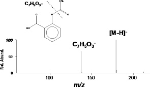
    above is mass spectrum of aspirin

    o-acetoxybenzoic acid
    C9H8O4              (Mass of molecular ion:    180)

     Source Temperature: 170 °C
       Sample Temperature: 100 °C
       DIRECT, 75 eV


    13 C NMR


    above is 13 C NMR OF ASPIRIN

    50.18 MHz
    C9 H8 O4 0.039 g : 0.5 ml CDCl3


     ppm   Int.  Assign.
    
          170.20   450      1
          169.76   510      2
          151.28   560      3
          134.90   924      4
          132.51  1000      5
          126.17   986      6
          124.01   974      7
          122.26   397      8
           20.99   674      9


    =================================================

    Animasi Kimia

    below is Raman spectra of aspirin


    NMR INTERPRETATIONS

    H-NMR spectral analysis
    Acetylsalicylic acid NMR spectra analysis, Chemical CAS NO. 50-78-2 NMR spectral analysis, Acetylsalicylic acid H-NMR spectrum
    CAS NO. 50-78-2, Acetylsalicylic acid H-NMR spectral analysis
    C-NMR spectral analysis
    Acetylsalicylic acid NMR spectra analysis, Chemical CAS NO. 50-78-2 NMR spectral analysis, Acetylsalicylic acid C-NMR spectrum
    CAS NO. 50-78-2, Acetylsalicylic acid C-NMR spectral analysis


    H, H-COSY spectrum

    In H, H-COSY spectrum are on both axes, the 1 H chemical shifts plotted; In principle, both the axes 1 to see H-NMR spectra. Thus, there is a symmetric to the diagonal diagram.
    1 H-NMR spectrum of acetylsalicylic acid

    Fig.2
    H, H-COSY spectrum of acetylsalicylic acid
    In the spectrum, only the range from 7.0 to 8.2 ppm is applied, because only here HH scalar couplings can be expected.

    Fig.3
    There are two types of signals:
    • Diagonal signals: join the coordinates δ a δ a (in core A), δ b δ b (in core B) ... on, but play no role in the evaluation of the couplings between different cores, since it is only the signal of a nucleus is. The diagonal with all its signals corresponding to the 1D H-NMR spectrum.
    EXAMPLE
    Acetylsalicylic acid
    7.13 ppm / 7.13 ppm = δ 2 δ 2 (H atom 2)
    7.34 ppm / 7.34 ppm = δ 4 δ 4 (H atom 4)
    7.61 ppm / 7.61 ppm = δ 3 δ 3 (H atom 3)
    8.11 ppm / 8.11 ppm = δ 5 δ 5 (H atom 5)
    • Cross signals: These signals are based on the scalar spin-spin coupling and are suitable for the evaluation of spectra of enormous importance.
    EXAMPLE
    Acetylsalicylic acid
    7.13 ppm / 7.61 ppm (δ 2 δ 3 ), and 7.61 ppm / 7.13 ppm (δ 3 δ 2 ) - vicinal coupling between the H-atoms 2 and 3
    7.34 ppm / 7.61 ppm (δ 4 δ 3 ) and 7.61 ppm / 7.34 ppm (δ 3 δ 4 ) - vicinal coupling between the H-atoms 4 and 3
    7.34 ppm / 8.11 ppm (δ 4 δ 5 ) and 8.11 ppm / 7.34 ppm (δ 5 δ 4 ) - vicinal coupling between the H-atoms 4 and 5
    In general it can be seen in the COSY spectrum each scalar coupling between two nuclei at four signals (two cross and two diagonal peaks) resulting connected a square; in the following example, the vicinal coupling between the H atoms is highlighted 3 and 4.

    Fig.4
    With good resolution of the COSY spectrum, the coupling constants can be determined from the fine structure of the cross and diagonal signals, but this is rarely done because of the 1-D H-NMR spectra is easily possible.
    DRUG APPROVALS BY DR ANTHONY MELVIN CRASTO .....FOR BLOG HOME CLICK HERE

    Join me on Linkedin

    View Anthony Melvin Crasto Ph.D's profile on LinkedIn

    Join me on Facebook FACEBOOK
    Join me on twitterFollow amcrasto on Twitter     
    Join me on google plus Googleplus

     amcrasto@gmail.com


    Khajuraho Group of Monuments is located in India
    Khajuraho Group of Monuments
    Location of Khajuraho Group of Monuments in India.

    Location in Madhya PradeshLocation in Madhya Pradesh

    1. Khajuraho Group of Monuments - Wikipedia, the free ...

      en.wikipedia.org/wiki/Khajuraho_Group_of_Monuments

      The Khajuraho Group of Monuments are a group of Hindu and Jain temples in Madhya Pradesh, India. About 620 kilometres (385 mi) southeast of New Delhi, ...


    Hotel Chandela - A Taj Leisure Hotel


    https://33.media.tumblr.com/1ccca0c990f7ce76796c2c2b35cb7f49/tumblr_noi3n89RZi1ur8m7ho1_500.gif