DR ANTHONY MELVIN CRASTO,WorldDrugTracker, helping millions, A 90 % paralysed man in action for you, I am suffering from transverse mylitis and bound to a wheel chair, With death on the horizon, nothing will not stop me except God................DR ANTHONY MELVIN CRASTO Ph.D ( ICT, Mumbai) , INDIA 25Yrs Exp. in the feld of Organic Chemistry,Working for GLENMARK GENERICS at Navi Mumbai, INDIA. Serving chemists around the world. Helping them with websites on Chemistry.Million hits on google, world acclamation from industry, academia, drug authorities for websites, blogs and educational contribution
Showing posts with label structure elucidation. Show all posts

Saturday, 31 October 2015

Computed NMR spectra predicts the structure of Nobilisitine A

Nobilisitine A was isolated by Evidente and coworkers, who proposed the structure 1.1 Banwell and co-workers then synthesized the enantiomer of 1, but its NMR did not correspond to that of reported for Nobilisitine A.; the largest differences are 4.7 ppm for the 13C NMR and 0.79 ppm for the 1H NMR.2

1
Lodewyk and Tantillo3 examined seven diastereomers of 1, all of which have a cis fusion between the saturated 5 and six-member rings (rings C and D). Low energy conformations were computed for each of these diasteromers at B3LYP/6-31+G(d,p). NMR shielding constants were then computed in solvent (using a continuum approach) at mPW1PW91/6-311+G(2d,p). A Boltzmann weighting of the shielding contants was then computed, and these shifts were then scaled as described by Jain, Bally and Rablen4 (discussed in this post). The computed NMR shifts for 1 were compared with the experimental values, and the mean deviations for the 13C and 1H svalues is 1.2 and 0.13 ppm, respectively. (The largest outlier is 3.4 ppm for 13C and 0.31 for 1H shifts.) Comparison was then made between the computed shifts of the seven diasteomers and the reported spectrum of Nobilisitine A, and the lowest mean deviations (1.4 ppm for 13C and 0.21 ppm for 1H) is for structure 2. However, the agreement is not substantially better than for a couple of the other diasteomers.

2
They next employed the DP4 analysis developed by Smith and Goodman5 for just such a situation – where you have an experimental spectrum and a number of potential diastereomeric structures. (See this post for a discussion of the DP4 method.)The DP4 analysis suggests that 2 is the correct structure with a probability of 99.8%.
Banwell has now synthesized the compound with structure 2 and its NMR matches that of the original natural product.6 Thus Nobilisitine A has the structure 2.

References

(1) Evidente, A.; Abou-Donia, A. H.; Darwish, F. A.; Amer, M. E.; Kassem, F. F.; Hammoda, H. A. m.; Motta, A., "Nobilisitine A and B, two masanane-type alkaloids from Clivia nobilis,"Phytochemistry, 1999, 51, 1151-1155, DOI: 10.1016/S0031-9422(98)00714-6.
(2) Schwartz, B. D.; Jones, M. T.; Banwell, M. G.; Cade, I. A., "Synthesis of the Enantiomer of the Structure Assigned to the Natural Product Nobilisitine A," Org. Lett., 2010, 12, 5210-5213, DOI:10.1021/ol102249q
(3) Lodewyk, M. W.; Tantillo, D. J., "Prediction of the Structure of Nobilisitine A Using Computed NMR Chemical Shifts," J. Nat. Prod., 2011, 74, 1339-1343, DOI: 10.1021/np2000446
(4) Jain, R.; Bally, T.; Rablen, P. R., "Calculating Accurate Proton Chemical Shifts of Organic Molecules with Density Functional Methods and Modest Basis Sets," J. Org. Chem., 2009, DOI:10.1021/jo900482q.
(5) Smith, S. G.; Goodman, J. M., "Assigning Stereochemistry to Single Diastereoisomers by GIAO NMR Calculation: The DP4 Probability," J. Am. Chem. Soc., 2010, 132, 12946-12959, DOI:10.1021/ja105035r
(6) Schwartz, B. D.; White, L. V.; Banwell, M. G.; Willis, A. C., "Structure of the Lycorinine Alkaloid Nobilisitine A," J. Org. Chem., 2011, ASAP, DOI: 10.1021/jo2016899


a tour

MYSORE, KARNATAKA, INDIA
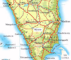
Map of mysore


////////////

1-Adamantyl cation – Predicting its NMR spectra

1-Adamantyl cation – Predicting its NMR spectra

What is required in order to compute very accurate NMR chemical shifts? Harding, Gauss and Schleyer take on the interesting spectrum of 1-adamantyl cation to try to discern the important factors in computing its 13C and 1H chemical shifts.1

1
To start, the chemical shifts of 1-adamtyl cation were computed at B3LYP/def2-QZVPP and
MP2/qz2p//MP2/cc-pVTZ. The root means square error (compared to experiment) for the carbon chemical shifts is large: 12.76 for B3LYP and 6.69 for MP2. The proton shifts are predicted much more accurately with an RMS error of 0.27 and 0.19 ppm, respectively.
The authors speculate that the underlying cause of the poor prediction is the geometry of the molecule. The structure of 1 was optimized at HF/cc-pVTZ, MP2/cc-pVTZ and CCSD(T)/pVTZ and then the chemical shifts were computed using MP2/tzp with each optimized geometry. The RMS error of the 12C chemical shifts are HF/cc-pVTZ: 9.55, MP2/cc-pVTZ: 5.62, and CCSD(T)/pVTZ: 5.06. Similar relationship is seen in the proton chemical shifts. Thus, a better geometry does seem to matter. The CCSD(T)/pVTZ optimized structure of 1 is shown in Figure 1.

1
Figure 1. CCSD(T)/pVTZ optimized structure of 1.
Unfortunately, the computed chemical shifts at CCSD(T)/qz2p//CCSD(T)/cc-pVTZ are still in error; the RMS is 4.78ppm for the carbon shifts and 0.26ppm for the proton shifts. Including a correction for the zero-point vibrational effects and adjusting to a temperature of 193 K to match the experiment does reduce the error; now the RMS for the carbon shifts is 3.85 ppm, with the maximum error of 6 ppm for C3. The RMS for the proton chemical shifts is 0.21ppm.
The remaining error they attribute to basis set incompleteness in the NMR computation, a low level treatment of the zero-point vibrational effects (which were computed at HF/tz2p), neglect of the solvent, and use of a reference in the experiment that was not dissolved in the same media as the adamantyl cation.
So, to answer our opening question – it appears that a very good geometry and treatment of vibrational effects is critical to accurate NMR shift computation of this intriguing molecule. Let the
computational chemist beware!

References

(1) Harding, M. E.; Gauss, J.; Schleyer, P. v. R., "Why Benchmark-Quality Computations Are Needed To Reproduce 1-Adamantyl Cation NMR Chemical Shifts Accurately," J. Phys. Chem. A, 2011, 115, 2340-2344, DOI: 10.1021/jp1103356


//////////

Welwitindolinones structure


A quick note here on the use of computed NMR to determine stereochemical structure. The Garg group synthesized two “oxidized welwitindolines”, compounds 1 and 2.1 The relative stereochemistry at the C3 position (the carbon with the hydroxy group) was unknown.

1

2
Low energy gas-phase conformers of both epimers of 1 and 2 were optimized at B3LYP/6-31+G(d,p). (These computations were done by the Tantillo group.) See Figure 1 for the optimized lowest energy conformers. Using these geometries the NMR chemical shifts were computed at mPW1PW91/6-311+G(d,p) with implicit solvent (chloroform). The chemical shifts were Boltzmann-weighted and scaled according to the prescription (see this post) of Jain, Bally and Rablen.2 The computed chemical shifts were then compared against the experimental NMR spectra. For both 1and 2, the 13C NMR shifts could not readily distinguish the two epimers. However, the computed 1H chemical shifts for the S epimer of each compound was significantly in better agreement with the experimental values; the mean average deviation for the S epimer of 2 is 0.08 ppm but 0.36ppm for the R epimer. As a check of these results, DP4 analysis3 (see this post) of 2 indicated a 100% probability for the S epimer using only the proton chemical shifts or with the combination of proton and carbon data.

1

2
Figure 1. B3LYP/6-31+G(d,p) optimized geometries of the
lowest energy conformations of 1 and 2.

References

(1) Quasdorf, K. W.; Huters, A. D.; Lodewyk, M. W.; Tantillo, D. J.; Garg, N. K., "Total Synthesis of Oxidized Welwitindolinones and (-)-N-Methylwelwitindolinone C Isonitrile," J. Am. Chem. Soc. 2011,134, 1396-1399, DOI: 10.1021/ja210837b
(2) Jain, R.; Bally, T.; Rablen, P. R., "Calculating Accurate Proton Chemical Shifts of Organic Molecules with Density Functional Methods and Modest Basis Sets," J. Org. Chem. 2009, 74, 4017-4023, DOI: 10.1021/jo900482q.
(3) Smith, S. G.; Goodman, J. M., "Assigning Stereochemistry to Single Diastereoisomers by GIAO NMR Calculation: The DP4 Probability," J. Am. Chem. Soc. 2010, 132, 12946-12959, DOI:10.1021/ja105035r


/////

Wednesday, 25 September 2013

VALSARTAN SPECTRAL DATA

 
VALSARTAN

mp 114–118 °C; 

1H NMR (400 MHz, DMSO-d6): δ 12.6 (brs, 1H), 7.72 (m, 4H), 7.24 (m, 1H), 7.15 (m, 2H), 6.94 (m, 1H), 4.58 (m, 1H), 4.40 (m, 1H), 3.33 (m, 1H), 2.25 (m, 1H), 1.52 (m, 6H), 0.9 (m, 3H), 0.84 (m, 3H), 0.74 (m, 3H); 


13C NMR (100 MHz, DMSO-d6): δ 174.0, 172.4, 171.8, 141.7, 138.2, 131.54, 131.1, 131.0, 129.3,128.8, 128.2, 127.4, 126.7, 70.3, 63.4, 49.9, 32.9, 28.05, 27.3, 22.2, 20.6, 14.2; 


ESIMS: m/z calcd [M]+: 435; found: 436 [M+H]+; HRMS (ESI): m/z calcd [M]+: 435.5187; found: 435.5125 [M]+


US 7439261 B2

1H-NMR (CDCl3) (0.80-1.15 (m, 9H); 1.20-1.50 (m, 2H); 1.60-1.80 (m, 2H); 2.60 (t, 2H); 2.65-2.80 (m, 2H), 3.70 (d, 1H), 4.10 (d, 0.3 H), 4.30 (d, 0.7 H), 4.90 (d, 0.7H), 5.2 (d, 0.3H); 7.00 (d, 0.3H); 7.10-7.20 (m, 4H), 7.40-7.60 (m, 3H), 7.85 (d, 0.7 H).


SHORT DESCRIPTION


Valsartan, N-(1-oxopentyl)-N-[[2′-(1H-tetrazol-5-yl)[1,1′-biphenyl]-4-yl]methyl]-L-valine, is a known anti-hypertensive agent having the following formula (I):
Figure US07439261-20081021-C00001
Valsartan and its preparation are disclosed in U.S. Pat. No. 5,399,578, in particular in Example 16. One of the synthetic routes according to U.S. Pat. No. 5,399,578 can be schematically represented as follows:
Figure US07439261-20081021-C00002
Figure US07439261-20081021-C00003
The synthetic pathway comprises various steps, among which:

    • coupling of compound (3) with 2-chlorobenzonitrile to obtain compound (4),
    • radicalic bromination of compound (4) to give compound (5),
    • transformation of the brominated derivative (5) into the respective aldehyde derivative (6),
    • reductive alkylation of compound (6) to obtain intermediate (8),
    • acylation of compound (8) to obtain intermediate (9),
    • conversion of the cyano group to the tetrazole group to afford intermediate (10),
    • deprotection of the carboxylic group by hydrogenolysis to obtain valsartan.
  • It is marketed as the free acid under the name DIOVAN. DIOVAN is prescribed as oral tablets in dosages of 40 mg, 80 mg, 160 mg and 320 mg ofvalsartan.
  • [0004]
    Valsartan and/or its intermediates are disclosed in various references, including: U.S. Pat. Nos. 5,399,578 ,5,965,592 5,260,325 6,271,375 WO 02/006253 WO 01/082858 WO 99/67231 WO 97/30036 , Peter Bühlmayer, et. al., Bioorgan. & Med. Chem. Let., 4(1) 29-34 (1994), Th. Moenius, et. al., J. Labelled Cpd. Radiopharm., 43(13) 1245 - 1252 (2000), and Qingzhong Jia, et. al., Zhongguo Yiyao Gongye Zazhi, 32(9) 385-387 (2001), all of which are incorporated herein by reference.
  • [0005]
    Valsartan is an orally active specific angiotensin II antagonist acting on the AT1 receptor subtype. Valsartan is prescribed for the treatment of hypertension. U.S. Pat. No. 6,395,728 is directed to use of valsartan for treatment of diabetes related hypertension. U.S. Pat. Nos. 6,465,502 and 6,485,745 are directed to treatment of lung cancer with valsartan. U.S. Pat. No. 6,294,197 is directed to solid oral dosage forms of valsartan
GOOD ARTICLES

http://users.uoa.gr/~tmavrom/2009/valsartan2009.pdf

http://www.acgpubs.org/JCM/2009/Volume%203/Issue%201/JCM-0908-14.pdf

https://www.beilstein-journals.org/bjoc/single/printArticle.htm?publicId=1860-5397-6-27 REPORTS
 mp 114–118 °C; 1H NMR (400 MHz, DMSO-d6): δ 12.6 (brs, 1H), 7.72 (m, 4H), 7.24 (m, 1H), 7.15 (m, 2H), 6.94 (m, 1H), 4.58 (m, 1H), 4.40 (m, 1H), 3.33 (m, 1H), 2.25 (m, 1H), 1.52 (m, 6H), 0.9 (m, 3H), 0.84 (m, 3H), 0.74 (m, 3H); 13C NMR (100 MHz, DMSO-d6): δ 174.0, 172.4, 171.8, 141.7, 138.2, 131.54, 131.1, 131.0, 129.3,128.8, 128.2, 127.4, 126.7, 70.3, 63.4, 49.9, 32.9, 28.05, 27.3, 22.2, 20.6, 14.2; ESIMS: m/z calcd [M]+: 435; found: 436 [M+H]+; HRMS (ESI): m/z calcd [M]+: 435.5187; found: 435.5125 [M]+


Valsartan 

Structural formula

UV - Spectrum


Conditions : Concentration - 1 mg / 100 ml
The solvent designation schedule
methanol 
water 
0.1Ðœ HCl 
0.1M NaOH 
maximum absorption249 nm250 nm248 nm251 nm
309302289311
e13400131001260013500

IR - spectrum

Wavelength (μm)
Wave number (cm -1 )

NMR spectrum


references


  • UV and IR Spectra. H.-W. Dibbern, R.M. Muller, E. Wirbitzki, 2002 ECV
  • NIST/EPA/NIH Mass Spectral Library 2008
  • Handbook of Organic Compounds. NIR, IR, Raman, and UV-Vis Spectra Featuring Polymers and Surfactants, Jr., Jerry Workman. Academic Press, 2000.
  • Handbook of ultraviolet and visible absorption spectra of organic compounds, K. Hirayama. Plenum Press Data Division, 1967.


Image result for VALSARTAN SYNTHESIS


CLIP

Image result for VALSARTAN SYNTHESIS


Scheme 2: (a) Et3N, CH2Cl2, 0 °C, 95%; (b) NaH, THF, 70%; (c) n-BuLi, 25 °C, THF, anhyd ZnCl2, −20 °C, Q-phos, Pd(OAc)2, 75 °C, 2 h, 80%; (d) 3 N NaOH, MeOH, reflux, 90%.

http://www.beilstein-journals.org/bjoc/single/articleFullText.htm?publicId=1860-5397-6-27

valsartan 8; mp 114–118 °C; 1H NMR (400 MHz, DMSO-d6): δ 12.6 (brs, 1H), 7.72 (m, 4H), 7.24 (m, 1H), 7.15 (m, 2H), 6.94 (m, 1H), 4.58 (m, 1H), 4.40 (m, 1H), 3.33 (m, 1H), 2.25 (m, 1H), 1.52 (m, 6H), 0.9 (m, 3H), 0.84 (m, 3H), 0.74 (m, 3H); 13C NMR (100 MHz, DMSO-d6): δ 174.0, 172.4, 171.8, 141.7, 138.2, 131.54, 131.1, 131.0, 129.3,128.8, 128.2, 127.4, 126.7, 70.3, 63.4, 49.9, 32.9, 28.05, 27.3, 22.2, 20.6, 14.2; ESIMS: m/z calcd [M]+: 435; found: 436 [M+H]+; HRMS (ESI): m/z calcd [M]+: 435.5187; found: 435.5125 [M]+


/////////